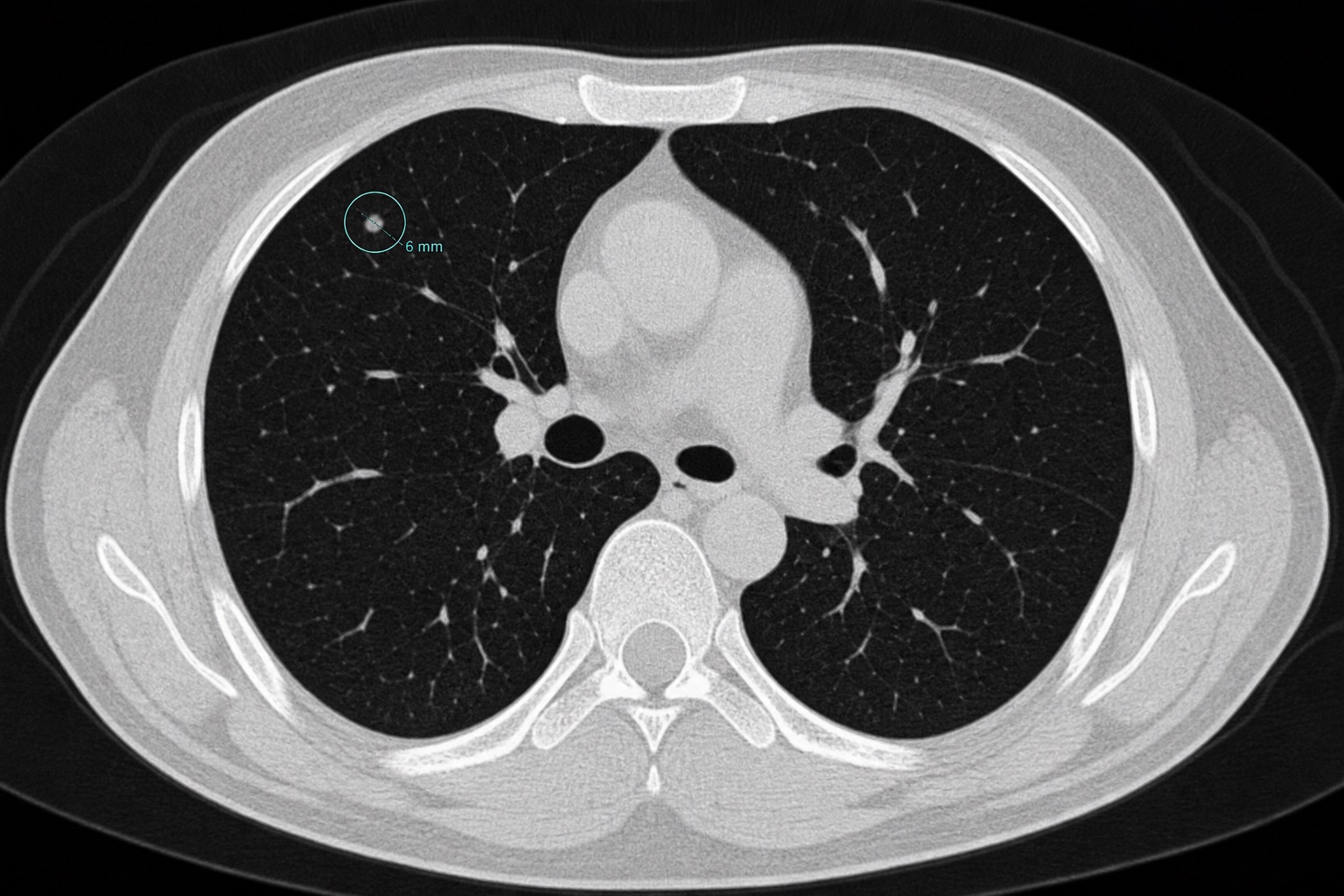
Understanding Findings

Lung Nodule Size Chart and When to Worry

A clear lung nodule size chart for CT findings: <4 mm, 4-6 mm, 6-8 mm, >8 mm, growth criteria, cancer risk features, and Fleischner follow-up.

CT Read Editorial10 min readPublished 2026-05-05

Reviewed by a board-certified radiologist

Lung Nodule Size Chart and When to Worry medical imaging guide cover
Radiologist reviewing chest CT lung window images with a pulmonary nodule
Synthetic editorial illustration for patient education. Not a real patient study or diagnosis.

Before You Read

Use this article as a plain-language guide, not a diagnosis

This guide explains common imaging terms, patterns, and follow-up questions so you can better understand your radiology report. Always confirm important findings with your radiologist or treating clinician.

Introduction

A lung nodule is a small, roughly round spot found on a chest X-ray or CT scan. Most nodules are benign — old infections, scars, or non-cancerous growths. Size is one of the most important features predicting whether a nodule needs follow-up. This guide explains the standard size thresholds and the widely-used Fleischner Society recommendations for solitary pulmonary nodules.

This lung nodule size chart is designed for people who saw a phrase like “4 mm pulmonary nodule,” “6 mm solid lung nodule,” or “8 mm ground-glass nodule” in a CT report and want to understand what size usually means. A lung nodule size chart helps organize risk, but size is only one part of lung nodule interpretation. Growth rate, density, margins, location, smoking history, and prior CT comparison are just as important.

Key takeaways from the lung nodule size chart

  • A lung nodule size chart is a starting point. Small nodules are usually low risk, but patient risk factors can change the follow-up plan.
  • Lung nodule size must be measured consistently. CT slice thickness, window settings, and measurement plane can affect the reported millimeters.
  • Lung nodule growth matters more than one measurement. A stable nodule over time is usually more reassuring than a single small measurement.
  • Lung nodule type changes follow-up. Solid, part-solid, and pure ground-glass nodules follow different surveillance rules.

1. Lung nodule size at a glance

Size (mm)Common interpretation
< 4 mmVery low risk — usually no follow-up in low-risk patients
4 - 6 mmLow risk — optional CT in 12 months for high-risk patients
6 - 8 mmIntermediate — CT in 6-12 months, then 18-24 months
> 8 mmHigher risk — consider PET-CT, biopsy, or 3-month CT follow-up

2. The Fleischner Society recommendations (2017)

The Fleischner Society guidelines stratify recommendations by:

  • Patient risk: low risk vs high risk (smokers, family history, COPD, exposure).
  • Nodule type: solid, part-solid (ground glass + solid), or pure ground glass.
  • Single vs multiple nodules.

Solid nodules < 6 mm in low-risk patients usually need no follow-up. Solid nodules > 8 mm warrant either short-interval CT, PET-CT, or biopsy depending on probability of malignancy.

3. Growth criteria

A nodule’s doubling time matters. Most malignancies double in volume within 30-400 days. Stability over 2 years for solid nodules (or 5 years for pure ground glass nodules) is generally reassuring. Volumetric measurement on serial CTs is more reliable than diameter alone.

Common lung nodule report phrases

A lung nodule report may say “solid lung nodule,” “ground-glass lung nodule,” “part-solid lung nodule,” “calcified granuloma,” or “stable pulmonary nodule.” Each phrase changes risk. A calcified lung nodule is often benign, while a part-solid lung nodule with a growing solid component needs closer follow-up. A stable lung nodule means the same spot has not grown compared with prior CT imaging.

When you use a lung nodule size chart, also read the surrounding language. A 5 mm lung nodule with smooth margins in a low-risk patient is different from a 5 mm lung nodule with spiculation in a smoker. Lung nodule follow-up depends on size, type, growth, patient risk, and whether previous imaging proves the lung nodule is unchanged.

4. Features that raise concern

  • Spiculated or irregular margins.
  • Upper lobe location (more common for lung cancer).
  • Solid or part-solid component > 6 mm.
  • Documented growth on serial scans.
  • Patient age > 50, current/former smoker, family history.

5. What to do next

Always follow your radiologist’s and pulmonologist’s recommendations. Bring previous imaging to follow-up appointments — comparing scans is more valuable than any single image. For rapid help understanding a CT scan that mentions a nodule, try our chest CT interpretation tool.

Use this lung nodule size chart to understand the language in your report, then ask your clinician whether the nodule is solid, part-solid, or ground-glass; whether it has grown; and whether the Fleischner Society follow-up schedule applies to your personal risk profile.

Frequently asked questions

Related articles