Einleitung
Ein CT-Scan (Computertomographie) setzt Hunderte von Röntgenbildern zu einer 3D-Karte Ihres Körpers zusammen. Für jemanden ohne medizinischen Hintergrund kann das Ergebnis einschüchternd wirken: Hunderte von grau-weißen Bildern in verschiedenen Ausrichtungen, mit kryptischen Beschriftungen wie „axial 2,5 mm“ oder „HU +45“.
Dieser Leitfaden bietet Ihnen eine praktische, anfängerfreundliche Einführung in das Lesen eines CT-Scans: Was die Ausrichtungen bedeuten, wie Hounsfield-Einheiten (HU) die Gewebedichte beschreiben, welche Strukturen Sie zuerst lernen sollten und welche Warnsignale Sie Ihrem Arzt mitteilen sollten. Nichts davon ersetzt einen Radiologiebericht – aber es wird Ihnen helfen, Ihren eigenen Scan mit viel mehr Vertrauen zu lesen.
Wenn Sie nach wie man einen CT-Scan liest gesucht haben, versuchen Sie wahrscheinlich, die Bilder auf Ihrem Bildschirm mit der Sprache in einem CT-Bericht zu verbinden. Dieser Artikel wiederholt bewusst die grundlegenden Schritte der CT-Scan-Interpretation: Identifizieren Sie die Ebene, wählen Sie das richtige Fenster, verstehen Sie die Hounsfield-Einheiten, vergleichen Sie beide Seiten und ordnen Sie dann den Befund dem Berichtseindruck zu. Dieser systematische Prozess des CT-Scan-Lesens verhindert, dass zufälliges Scrollen verwirrend oder irreführend wird.
Wichtige Erkenntnisse vor dem Lesen eines CT-Scans
- Die CT-Scan-Interpretation beginnt mit der Orientierung. Axiale, koronale und sagittale Bilder beantworten unterschiedliche anatomische Fragen.
- Die CT-Scan-Dichte wird in Hounsfield-Einheiten gemessen. Luft, Fett, Flüssigkeit, Weichgewebe, Blut, Knochen und Metall haben jeweils erkennbare HU-Bereiche.
- Die Einstellungen des CT-Scan-Fensters sind wichtig. Lungenfenster, Knochenfenster, Gehirnfenster und Weichgewebefenster können dieselbe Krankheit offensichtlich oder nahezu unsichtbar erscheinen lassen.
- CT-Scan-Befunde benötigen klinischen Kontext. Ein Radiologe vergleicht Symptome, Laborergebnisse, frühere Bildgebung und Kontrastphase, bevor er eine Bedeutung zuweist.
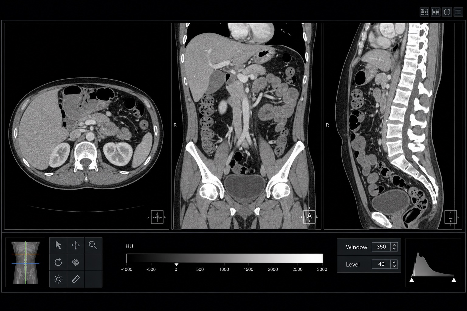
1. Orientieren Sie sich: axial, koronal, sagittal
Die meisten CT-Viewer zeigen drei orthogonale Serien:
- Axial – Schnitte von Kopf bis Fuß, als ob man von den Füßen nach oben schaut. Die rechte Körperseite erscheint auf der linken Seite des Bildes.
- Koronal – Schnitte von vorne nach hinten, als ob man eine Person frontal betrachtet.
- Sagittal – Schnitte von Seite zu Seite, als ob man jemanden im Profil betrachtet.
Bestätigen Sie immer die Orientierungsbeschriftungen (R für rechts, L für links, A für anterior, P für posterior). Auf axialen Bildern befindet sich die rechte Seite des Patienten konventionell auf Ihrer linken Seite.
2. Lesen Sie die „Farbe“ – Hounsfield-Einheiten (HU)
CT misst die Gewebedichte relativ zu Wasser. Jeder Pixel hat einen Hounsfield-Einheitenwert:
- Luft: etwa −1000 HU (schwarz)
- Fett: etwa −100 HU
- Wasser: 0 HU
- Weichgewebe (Muskel, Organe): +30 bis +60 HU
- Akutes Blut: +50 bis +90 HU (heller als Gehirn bei einem Kopf-CT)
- Knochen: +400 bis +1000 HU (sehr weiß)
- Metall/Kontrastmittel: weit über +1000 HU
Das Anpassen von Fenster/Pegel hebt verschiedene Gewebe hervor. Ein „Lungenfenster“ betont Luft, ein „Knochenfenster“ betont verkalkte Strukturen und ein „Weichgewebefenster“ ist am besten für Organe geeignet.
Häufige Formulierungen in CT-Berichten
Ein CT-Bericht verwendet oft wiederholte Kurzformen, die leichter zu verstehen sind, sobald man das Muster kennt. „Keine akute CT-Anomalie“ bedeutet normalerweise, dass der CT-Scan keinen dringenden Befund zeigte. „Klinisch korrelieren“ bedeutet, dass der CT-Scan-Befund mit Symptomen oder Laborergebnissen abgeglichen werden muss. „Vergleich mit früherem CT-Scan“ bedeutet, dass der Radiologe überprüft hat, ob der Befund neu, stabil, größer oder kleiner ist. „Eingeschränkter CT-Scan aufgrund von Bewegung“ bedeutet, dass Unschärfe das Vertrauen in die CT-Scan-Interpretation verringert hat.
Beim Lesen eines CT-Scans sollten Sie immer trennen, was der CT-Scan klar zeigt, von dem, was der CT-Scan nicht beantworten kann. Ein CT-Scan ist hervorragend geeignet für akute Blutungen, Frakturen, Nierensteine, Lungenknötchen, Darmverschluss und viele Krebsarten, aber der CT-Scan kann frühe Infektionen, kleine Weichteilverletzungen oder Krankheiten übersehen, die eine MRT oder Ultraschall erfordern.
Bei der praktischen CT-Scan-Interpretation behandeln Sie den CT-Scan als eine strukturierte Karte: Scannen Sie die Knochen, scannen Sie die Organe, scannen Sie die Gefäße und scannen Sie die Weichteile jedes Mal in derselben Reihenfolge.
Eine konsistente CT-Scan-Checkliste verwandelt das CT-Scan-Lesen in einen wiederholbaren CT-Scan-Interpretationsprozess anstatt in eine Ratespiel.
3. Systematisch lesen: ein „ABCDE“ für den Körper
Wählen Sie eine konsistente Reihenfolge. Eine einfache Körper-CT-Routine:
- Anatomie zuerst – bestätigen Sie, dass der abgebildete Bereich der Anforderung entspricht.
- Knochen – scrollen Sie das Knochenfenster nach Frakturen, Läsionen.
- Herz-Lunge – Herzgröße, Mediastinum, Lunge.
- Verdauungs-/Bauchorgane – Leber, Milz, Bauchspeicheldrüse, Nieren, Darm.
- Alles andere – Gefäße, Lymphknoten, Weichteile.
4. Mit oder ohne Kontrastmittel?
Jodhaltiges Kontrastmittel hilft, Blutgefäße, Nieren und viele Tumore hervorzuheben. Ihr Scan kann mehrere Phasen haben: arteriell, portalvenös, verzögert. Berichte geben normalerweise an, in welcher Phase ein Befund am besten sichtbar ist. Reaktionen auf Kontrastmittel sind selten, aber möglich – informieren Sie das Team immer über Nierenprobleme oder frühere Reaktionen.
5. Warnsignale, die Sie Ihrem Arzt mitteilen sollten
- Freie Luft außerhalb des Darms (deutet auf Perforation hin).
- Akutes Blut (hohe HU) im Gehirn oder Bauchraum.
- Lungenembolie: Füllungsdefekte in den Lungenarterien bei der CT-Angiographie.
- Lungenknötchen > 6 mm (insbesondere bei Rauchern).
- Aortenaneurysma oder -dissektion.
Auch wenn Sie etwas Auffälliges entdecken können, ist der Radiologiebericht das, was die Behandlung leitet. Verwenden Sie diesen Leitfaden, um bessere Fragen zu stellen.
Nächste Schritte
Versuchen Sie, Ihren Scan in unser KI-CT-Analyse-Tool hochzuladen, um eine sofortige, leicht verständliche Zusammenfassung zu erhalten, oder öffnen Sie ihn in unserem kostenlosen DICOM-Viewer, um die Schnitte selbst zu scrollen.
Der beste Weg, wie man einen CT-Scan liest, ist, die ursprüngliche DICOM-Datei zu öffnen, langsam durch jede Ebene zu scrollen und Ihre Beobachtungen mit dem offiziellen Bericht zu vergleichen. Verwenden Sie diesen CT-Scan-Leseleitfaden als Checkliste, nicht als Diagnose.
Frequently asked questions
Nützliche Tools
Verwandte Artikel
So lesen Sie einen MRT-Scan
Lernen Sie, wie man einen MRT-Scan in einfacher Sprache liest: T1 vs. T2, FLAIR, DWI/ADC, Kontrastmittelanreicherung, Bildebenen, Artefakte und Berichtsterminologie.
So lesen Sie eine Röntgenaufnahme des Brustkorbs
Lernen Sie, wie man eine Röntgenaufnahme des Brustkorbs mit einer systematischen ABCDE-Methode liest: Bildqualität, Atemwege, Lunge, Herzgröße, Zwerchfell, häufige Anomalien und Warnzeichen.
Tabelle zur Größe von Lungenrundherden und wann man sich Sorgen machen sollte
Eine klare Tabelle zur Größe von Lungenrundherden für CT-Befunde: <4 mm, 4-6 mm, 6-8 mm, >8 mm, Wachstumskriterien, Merkmale des Krebsrisikos und Fleischner-Nachsorge.