مدونة CT Read
تنشر مدونة CT Read أدلة تفسير التصوير الطبي للمرضى والعائلات والطلاب والأطباء الذين يرغبون في الحصول على تفسيرات أوضح لنتائج التصوير المقطعي المحوسب والتصوير بالرنين المغناطيسي والأشعة السينية للصدر والموجات فوق الصوتية وأدوات DICOM وتقارير الأشعة.
يركز كل دليل على سؤال عملي واحد: كيفية قراءة التصوير المقطعي المحوسب، وكيفية قراءة التصوير بالرنين المغناطيسي، وماذا تعني البقع البيضاء في التصوير بالرنين المغناطيسي للدماغ، وكيف يؤثر حجم العقيدات الرئوية على المتابعة، أو كيف يظهر تمزق الغضروف الهلالي في التصوير بالرنين المغناطيسي للركبة. الهدف ليس استبدال طبيب الأشعة، بل مساعدتك على فهم الكلمات والصور وتوصيات المتابعة في تقريرك.
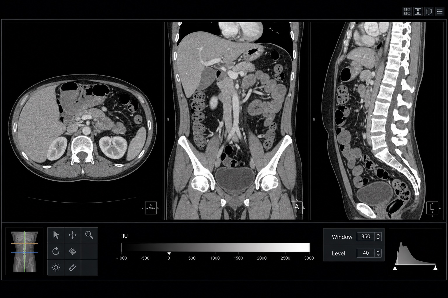
كيفية قراءة التصوير المقطعي المحوسب: دليل المبتدئين
تعلم كيفية قراءة التصوير المقطعي المحوسب خطوة بخطوة: الشرائح المحورية، والمناظر الإكليلية والساجيتالية، ووحدات هاونسفيلد، وإعدادات نافذة التصوير المقطعي المحوسب، والتشريح الرئيسي، والعلامات الحمراء العاجلة.
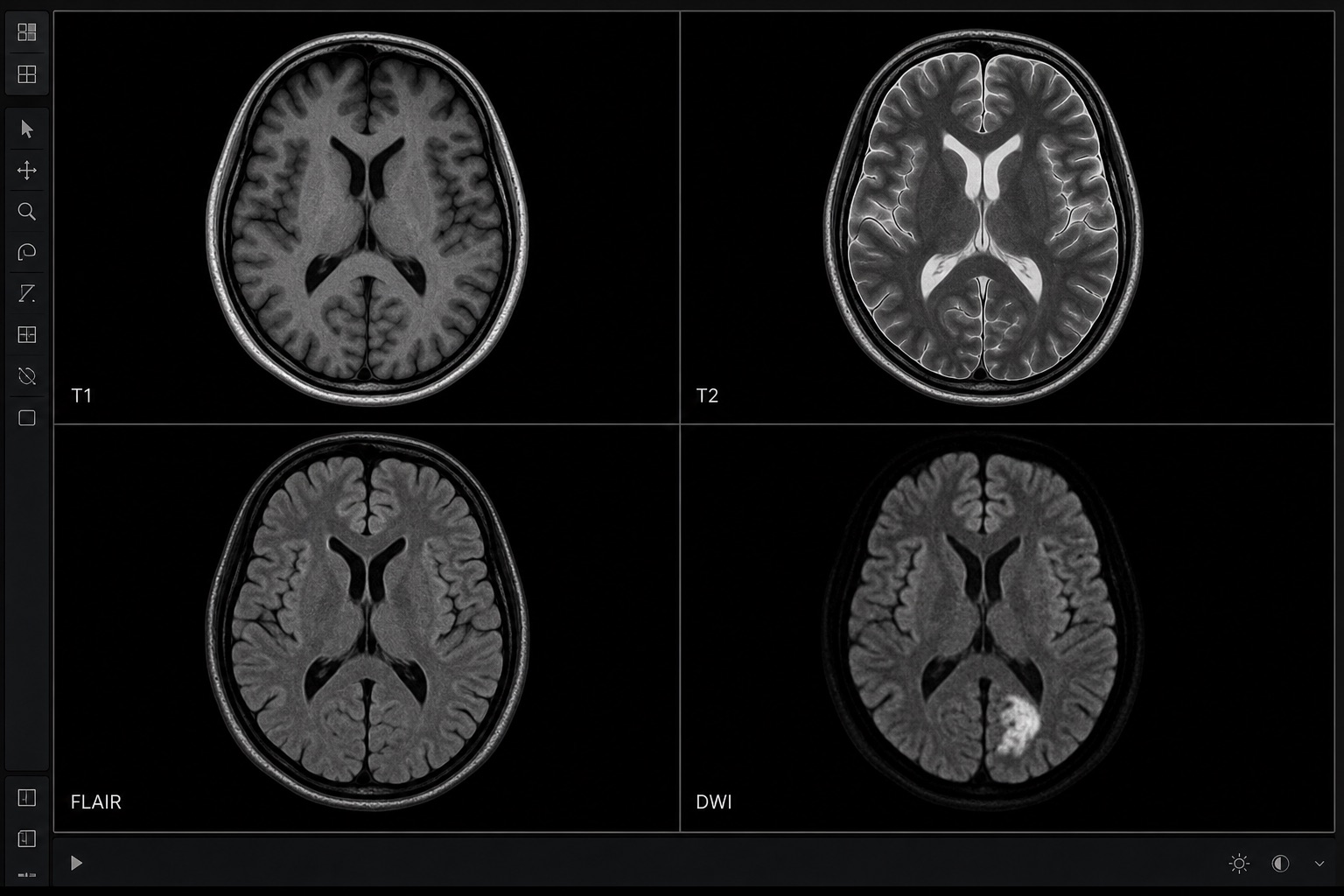
كيفية قراءة التصوير بالرنين المغناطيسي
تعلم كيفية قراءة التصوير بالرنين المغناطيسي بلغة مبسطة: T1 مقابل T2، FLAIR، DWI/ADC، تعزيز التباين، مستويات الصورة، القطع الأثرية، ومصطلحات التقرير.
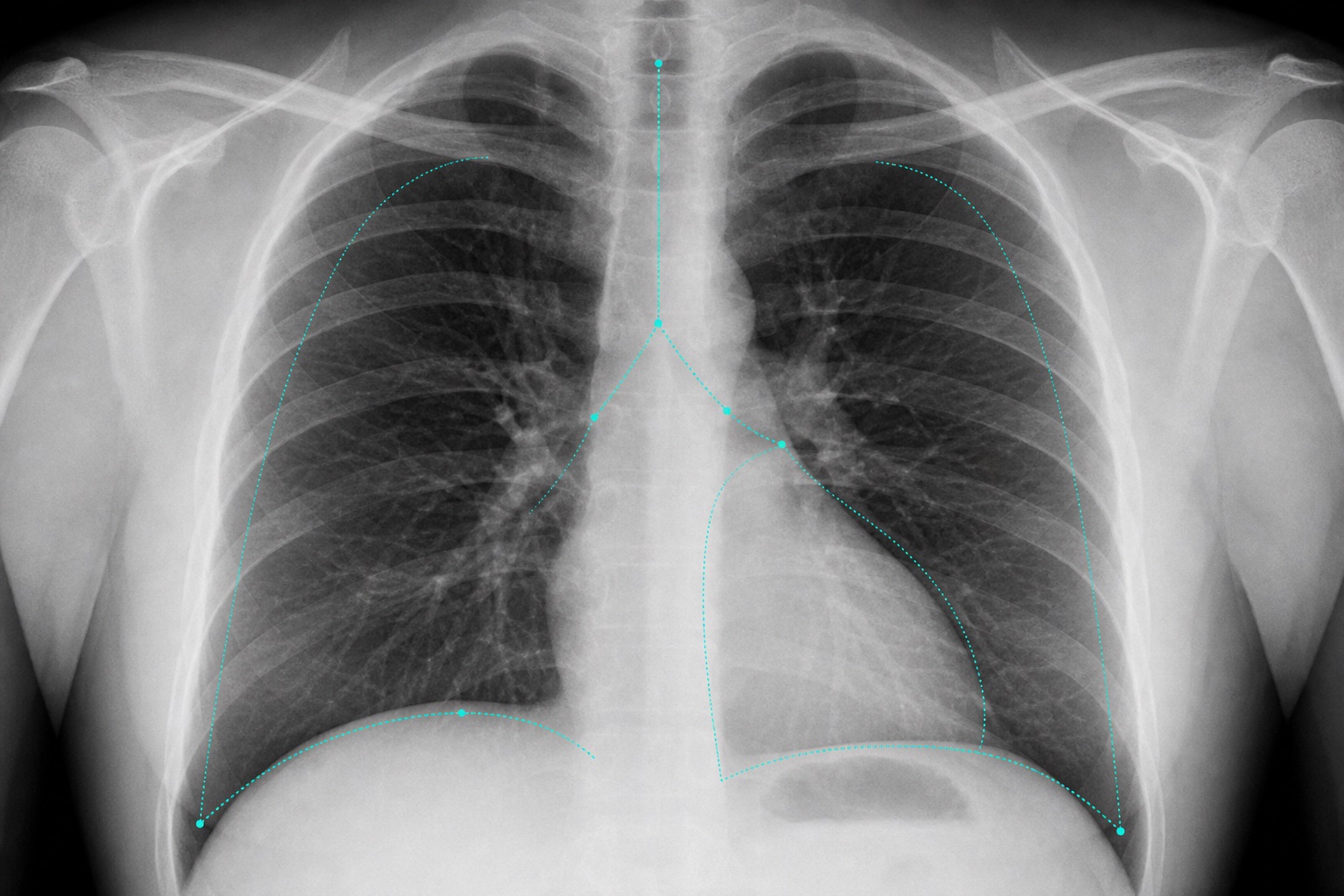
كيفية قراءة الأشعة السينية للصدر
تعلم كيفية قراءة الأشعة السينية للصدر باستخدام طريقة ABCDE المنهجية: جودة الصورة، مجرى الهواء، الرئتين، حجم القلب، الحجاب الحاجز، التشوهات الشائعة، والعلامات الحمراء.
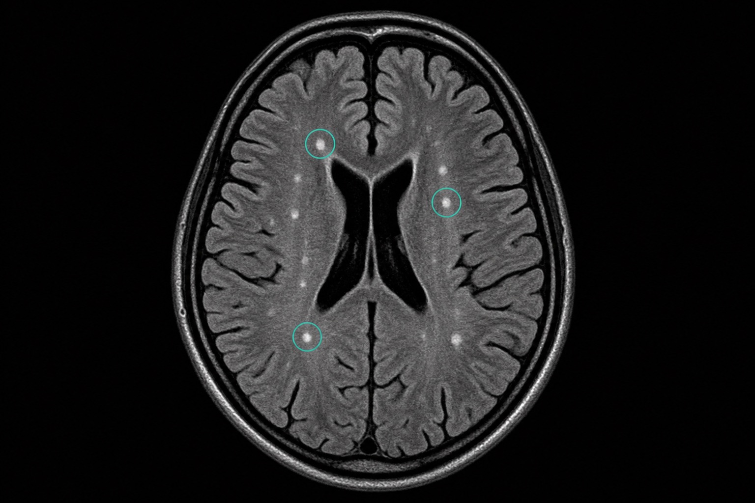
البقع البيضاء في التصوير بالرنين المغناطيسي للدماغ: ماذا تعني
فهم البقع البيضاء في التصوير بالرنين المغناطيسي للدماغ: فرط إشارات المادة البيضاء T2/FLAIR، بقع الصداع النصفي، مرض الأوعية الدموية الصغيرة، أنماط التصلب المتعدد، عوامل الخطر، والمتابعة.
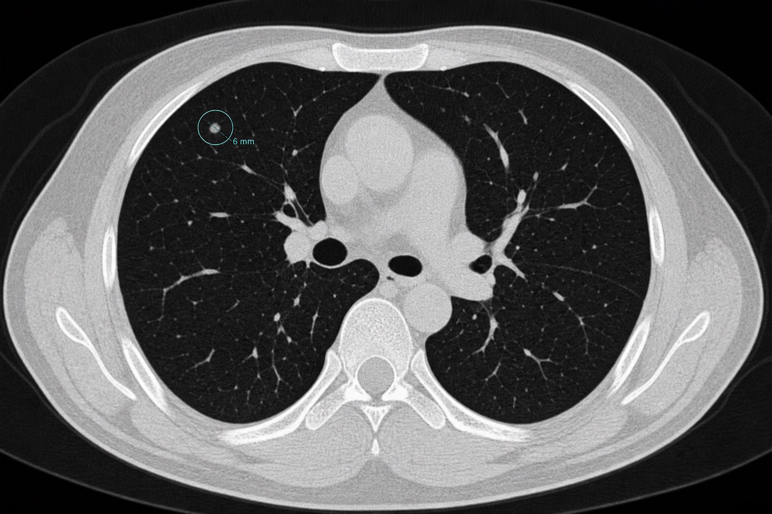
مخطط حجم العقيدات الرئوية ومتى يجب القلق
مخطط واضح لحجم العقيدات الرئوية لنتائج التصوير المقطعي المحوسب: <4 مم، 4-6 مم، 6-8 مم، >8 مم، معايير النمو، ميزات خطر السرطان، ومتابعة فليشنر.
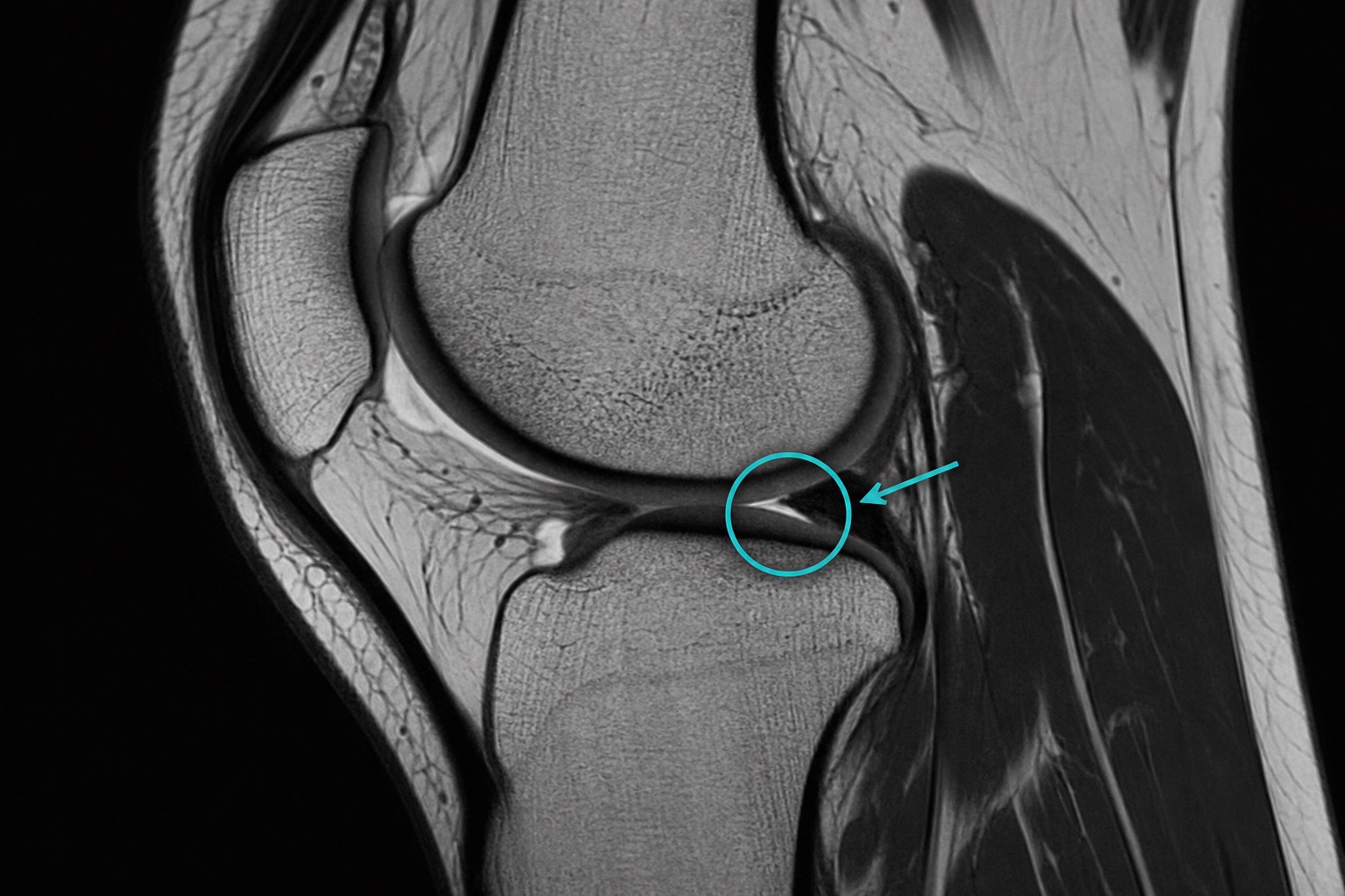
تمزق الغضروف الهلالي في التصوير بالرنين المغناطيسي: كيفية التعرف عليه
تعلم كيف يظهر تمزق الغضروف الهلالي في التصوير بالرنين المغناطيسي: إشارة تمزق الغضروف الهلالي، تمزقات القرن الخلفي، علامات تمزق مقبض الدلو، الدرجات، وأدلة العلاج.
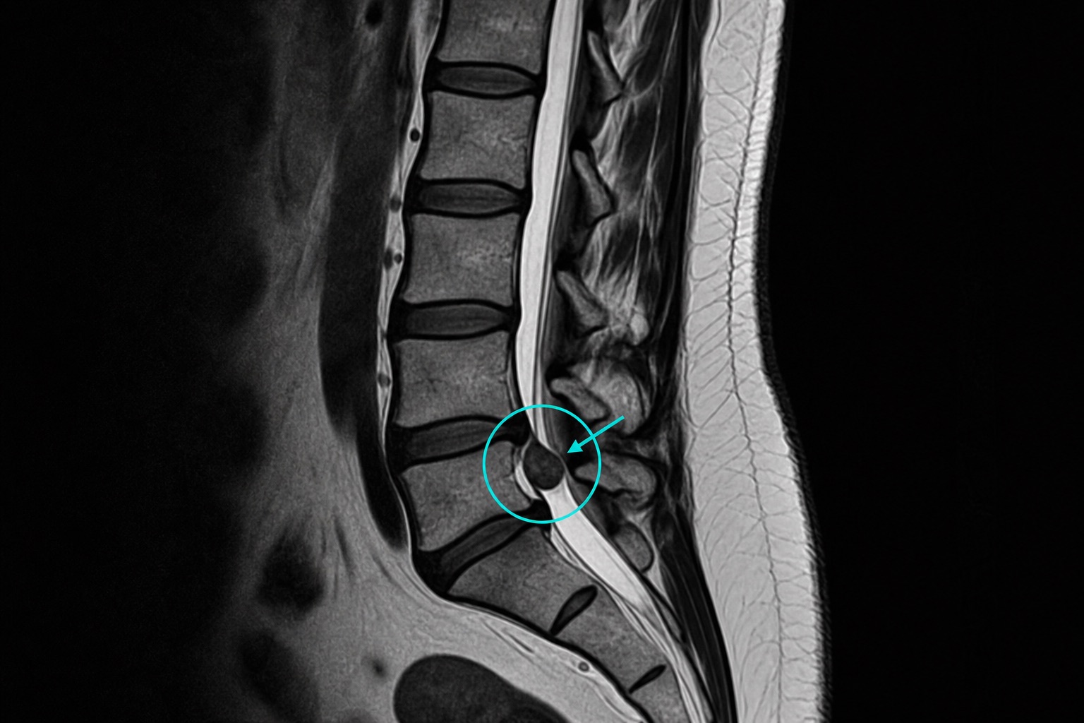
انزلاق القرص الغضروفي في التصوير بالرنين المغناطيسي: نصائح للقراءة
فهم انزلاق القرص الغضروفي في التصوير بالرنين المغناطيسي: انتفاخ القرص مقابل بروز القرص مقابل انزلاق القرص، مستويات التصوير بالرنين المغناطيسي للعمود الفقري القطني، انضغاط جذر العصب، تضيق العمود الفقري، والعلامات الحمراء.
مدونة CT Read
افهم الصور الطبية قبل موعدك القادم
تُنظم مدونة CT Read حول مواضيع بحثية عالية الأهمية: تفسير التصوير المقطعي المحوسب، تفسير التصوير بالرنين المغناطيسي، تفسير الأشعة السينية للصدر، متابعة العقيدات الرئوية، نتائج المادة البيضاء في التصوير بالرنين المغناطيسي للدماغ، تمزقات الغضروف الهلالي في التصوير بالرنين المغناطيسي للركبة، وانزلاق القرص الغضروفي في التصوير بالرنين المغناطيسي للعمود الفقري القطني. تشرح أدلة تفسير التصوير الطبي هذه كيف تبدو النتيجة، وما هي المشاكل الجسدية التي يمكن أن تشير إليها، ومتى تكون عاجلة، وما هو اختبار المتابعة الذي يُعتبر عادةً.
تفسير التصوير المقطعي المحوسب
تعلم اتجاه التصوير المقطعي المحوسب، ووحدات هاونسفيلد، ومراحل التباين، وعتبات حجم العقيدات الرئوية، والنتائج الخطيرة التي يجب مناقشتها مع الطبيب.
تفسير التصوير بالرنين المغناطيسي
تعلم تسلسلات التصوير بالرنين المغناطيسي مثل T1 و T2 و FLAIR و DWI والتصوير بالرنين المغناطيسي المعزز بالتباين، بما في ذلك كيفية وصف نتائج التصوير بالرنين المغناطيسي للدماغ والركبة والعمود الفقري القطني.
تفسير الأشعة السينية
استخدم طرق قراءة الأشعة السينية المنهجية للأشعة السينية للصدر، والأشعة السينية للعظام، والنتائج الطارئة مثل استرواح الصدر، والكسور، والانصباب، والتكثف.
DICOM وأدوات التصوير
افتح صور DICOM، وافحص البيانات الوصفية، وحوّل DICOM إلى JPG أو فيديو، وقم بإعداد ملفات التصوير للآراء الثانية، أو التدريس، أو البحث، أو تحليل الذكاء الاصطناعي.
كيفية استخدام أدلة الأشعة هذه
ابدأ بالمقالة التي تتطابق مع العبارة الدقيقة في تقرير الأشعة الخاص بك. إذا كان تقريرك يقول "فرط إشارات المادة البيضاء"، فاقرأ دليل البقع البيضاء في التصوير بالرنين المغناطيسي للدماغ. إذا كان يقول "عقيدة رئوية صلبة 6 مم"، فاستخدم مخطط حجم العقيدات الرئوية. إذا كان يقول "تمزق الغضروف الهلالي في القرن الخلفي"، فاقرأ دليل التصوير بالرنين المغناطيسي للغضروف الهلالي الممزق. ثم قارن المقالة بتقريرك الرسمي واطرح أسئلة محددة على طبيب الأشعة، أو طبيب الرعاية الأولية، أو أخصائي أمراض الرئة، أو طبيب الأعصاب، أو جراح العظام، أو أخصائي العمود الفقري.