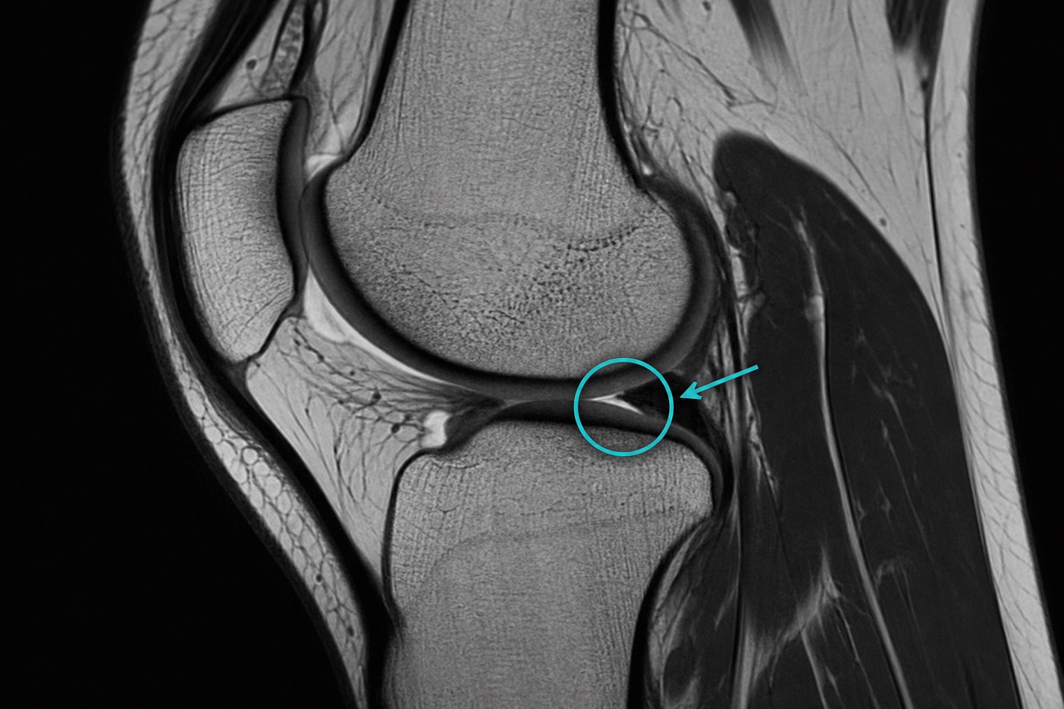
Introduction
The meniscus is the C-shaped cartilage that cushions and stabilizes the knee. Tears are common from sports, twisting injuries, or simple wear and tear with age. MRI is the gold standard test for diagnosing meniscal tears. This guide explains the common types of tears, how they appear on MRI, and what your report likely means.
If your report says torn meniscus on MRI, the key question is not just whether a tear exists, but which meniscus is torn, which part is involved, whether the tear reaches an articular surface, and whether the fragment is displaced. A torn meniscus on MRI can look very different depending on whether the tear is horizontal, radial, vertical, bucket-handle, root, or complex.
Key takeaways about a torn meniscus on MRI
- A torn meniscus on MRI usually shows bright signal reaching the surface. Grade 3 signal is more suspicious than internal degeneration.
- A torn meniscus on MRI is described by location. Medial vs lateral meniscus, anterior horn vs body vs posterior horn, and root involvement all matter.
- A torn meniscus on MRI is described by pattern. Bucket-handle and root tears are more likely to cause mechanical symptoms or require surgery.
- A torn meniscus on MRI must match symptoms. Many degenerative meniscal tears are incidental, especially in adults with osteoarthritis.
1. Quick anatomy review
- Two menisci per knee: medial (inner side, more commonly torn) and lateral (outer side).
- Each meniscus has an anterior horn, body, and posterior horn.
- The peripheral one-third has a blood supply (the “red zone”) and may heal; the inner two-thirds is mostly avascular.
2. How tears look on MRI
On sagittal proton-density and T2-weighted images, the meniscus appears as a uniformly dark wedge. A tear is suggested when an abnormal high-signal line:
- Reaches the articular surface (superior or inferior).
- Or distorts the meniscus shape.
High signal that does not touch the surface usually represents intrasubstance degeneration, not a true tear.
3. Common tear types
- Horizontal cleavage: a horizontal split, often degenerative, in older patients.
- Vertical longitudinal: traumatic, may evolve into a bucket-handle tear.
- Bucket-handle: a displaced fragment hanging into the joint, often locking the knee.
- Radial: a transverse tear from the inner edge outward; can disrupt circumferential fibers.
- Complex: combination of patterns, common with chronic tears.
4. Will I need surgery?
Not all meniscal tears need surgery. Decision factors include:
- Patient age and activity level.
- Tear type, location (red vs white zone), and size.
- Mechanical symptoms (locking, catching).
- Failure of conservative management (rest, PT, anti-inflammatories).
Many degenerative tears respond well to physical therapy; traumatic tears with mechanical symptoms in younger patients are more likely to need arthroscopy.
5. Grades of meniscal signal change
- Grade 1: punctate intrasubstance signal — degeneration, no tear.
- Grade 2: linear intrasubstance signal — degeneration, no tear.
- Grade 3: signal reaching the articular surface — true tear.
Common meniscus MRI report phrases
A meniscus MRI report may mention “posterior horn medial meniscus tear,” “horizontal meniscus tear,” “complex meniscus tear,” “meniscus root tear,” or “macerated meniscus.” These phrases describe the part of the meniscus involved and the tear pattern. A meniscus root tear can behave like loss of the entire meniscus cushion, while a small degenerative meniscus signal may not explain knee pain by itself.
Meniscus MRI interpretation should also look for associated findings: cartilage loss, bone marrow edema, ACL tear, MCL sprain, joint effusion, Baker cyst, and osteoarthritis. A meniscus tear with locking symptoms is interpreted differently from a meniscus tear discovered incidentally during imaging for chronic arthritis.
For a personalized AI explanation of your knee MRI, try the knee MRI interpretation tool.
When discussing a torn meniscus on MRI with a doctor, ask whether the tear is stable, displaced, repairable, and located in the red zone or white zone. Those details usually matter more than the word “tear” alone.
Frequently asked questions
Related articles
How to Read a CT Scan: A Beginner’s Guide
Learn how to read a CT scan step by step: axial slices, coronal and sagittal views, Hounsfield units, CT window settings, key anatomy, and urgent red flags.
How to Read an MRI Scan
Learn how to read an MRI scan in plain language: T1 vs T2, FLAIR, DWI/ADC, contrast enhancement, image planes, artifacts, and report terminology.
How to Read a Chest X-Ray
Learn how to read a chest X-ray using a systematic ABCDE method: image quality, airway, lungs, heart size, diaphragm, common abnormalities, and red flags.


