CT Read Blog
CT Read Blog publishes medical imaging interpretation guides for patients, families, students, and clinicians who want clearer explanations of CT scan, MRI scan, chest X-ray, ultrasound, DICOM tools, and radiology report findings.
Each guide focuses on one practical question: how to read a CT scan, how to read an MRI, what white spots on brain MRI mean, how lung nodule size changes follow-up, or how a torn meniscus appears on knee MRI. The goal is not to replace a radiologist, but to help you understand the words, images, and follow-up recommendations in your report.
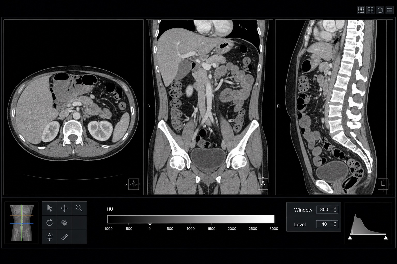
How to Read a CT Scan: A Beginner's Guide
Learn how to read a CT scan step by step: axial slices, coronal and sagittal views, Hounsfield units, CT window settings, key anatomy, and urgent red flags.
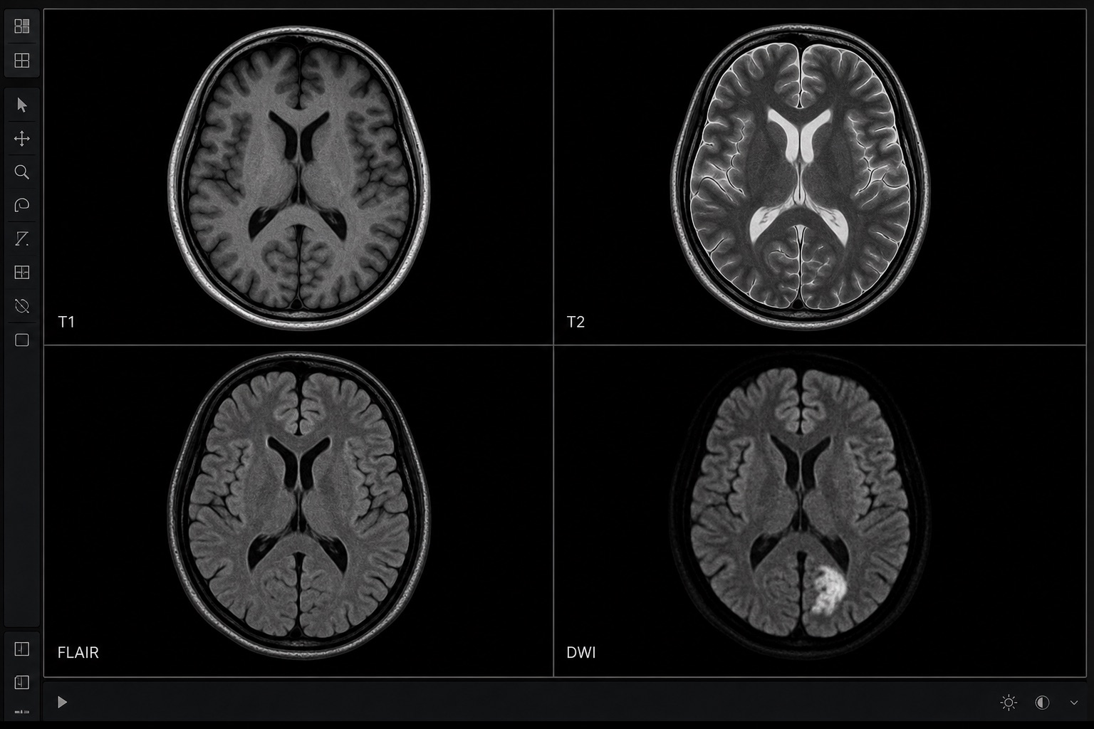
How to Read an MRI Scan
Learn how to read an MRI scan in plain language: T1 vs T2, FLAIR, DWI/ADC, contrast enhancement, image planes, artifacts, and report terminology.
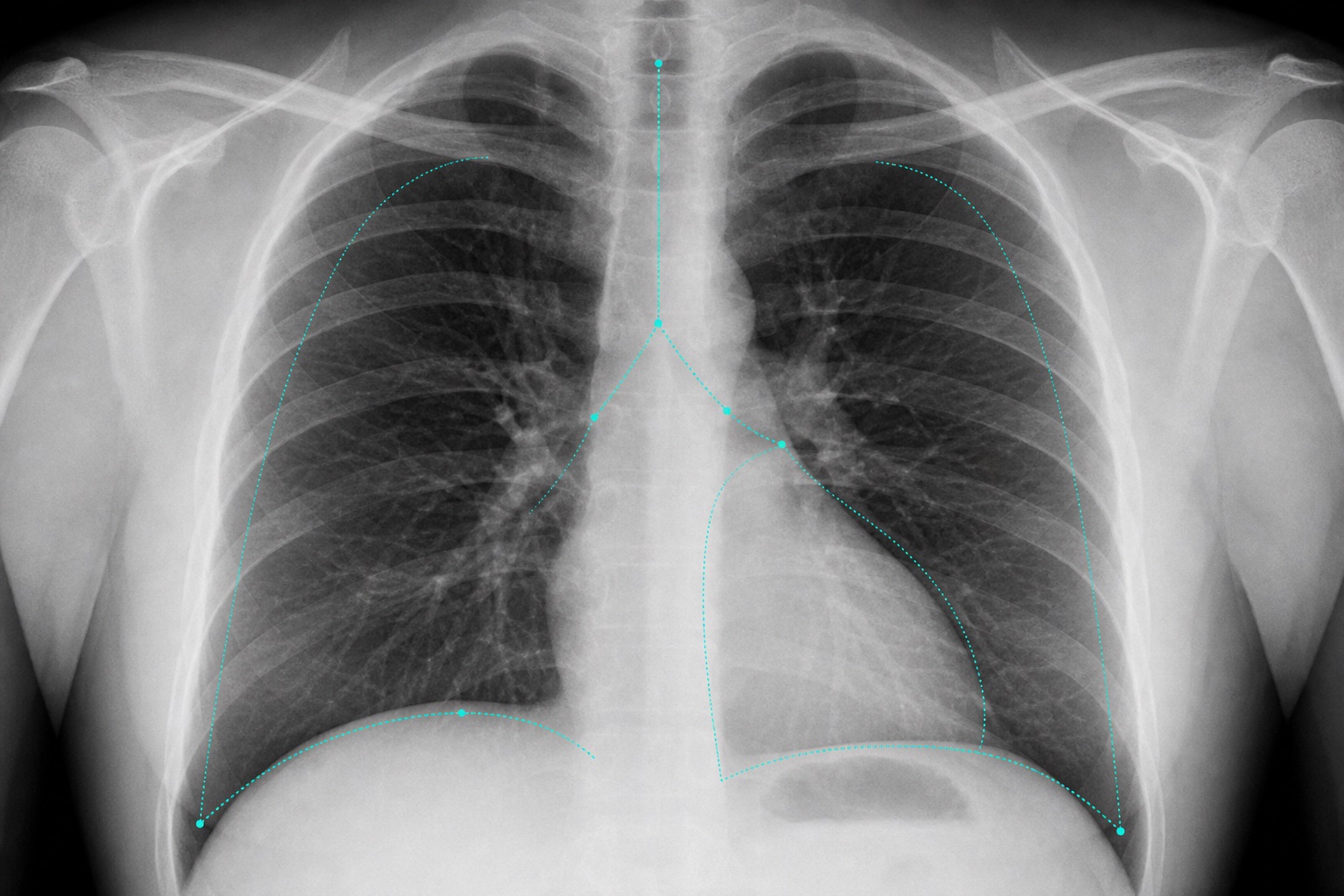
How to Read a Chest X-Ray
Learn how to read a chest X-ray using a systematic ABCDE method: image quality, airway, lungs, heart size, diaphragm, common abnormalities, and red flags.
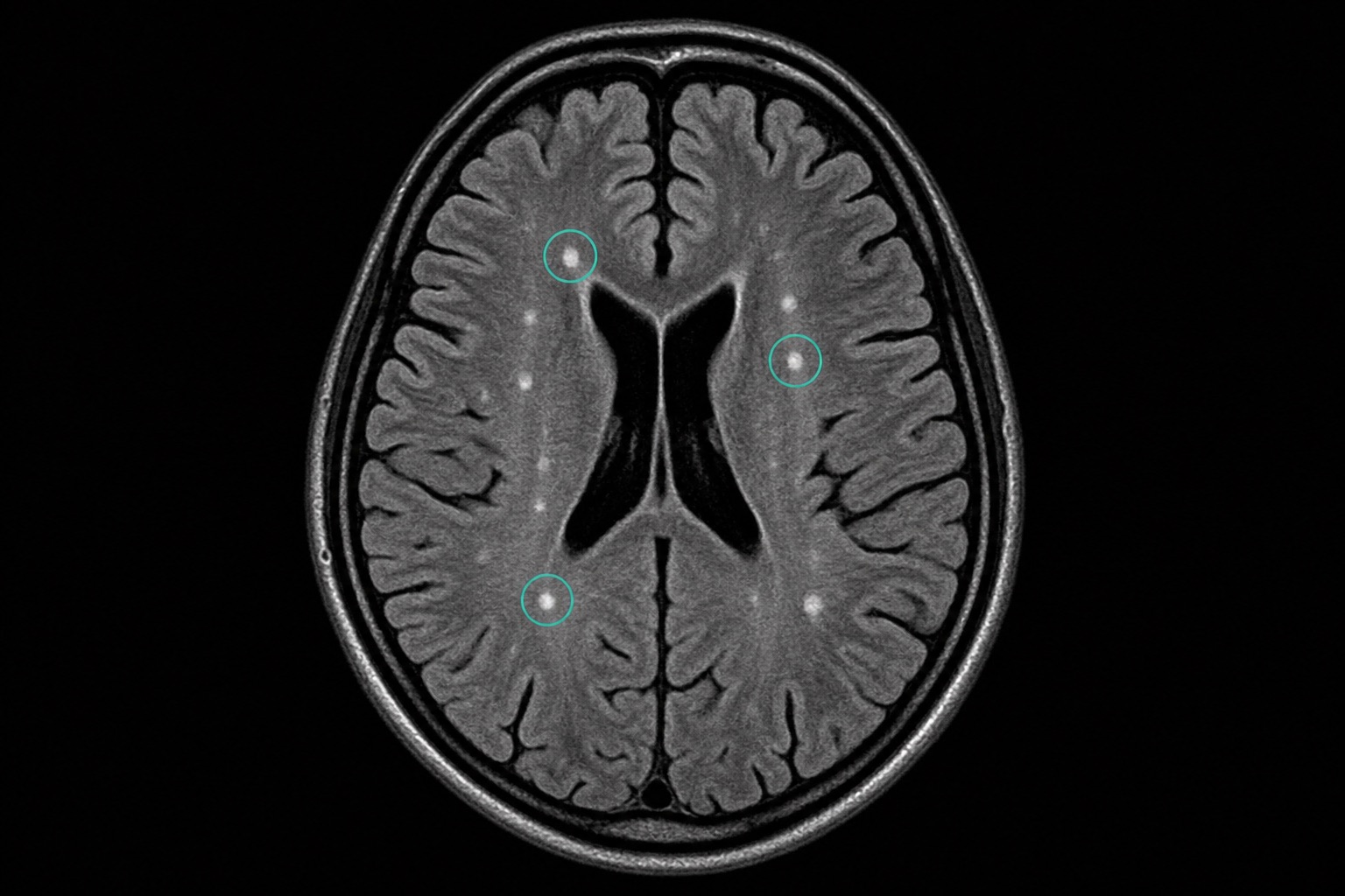
White Spots on Brain MRI: What They Mean
Understand white spots on brain MRI: T2/FLAIR white matter hyperintensities, migraine spots, small vessel disease, MS patterns, risk factors, and follow-up.
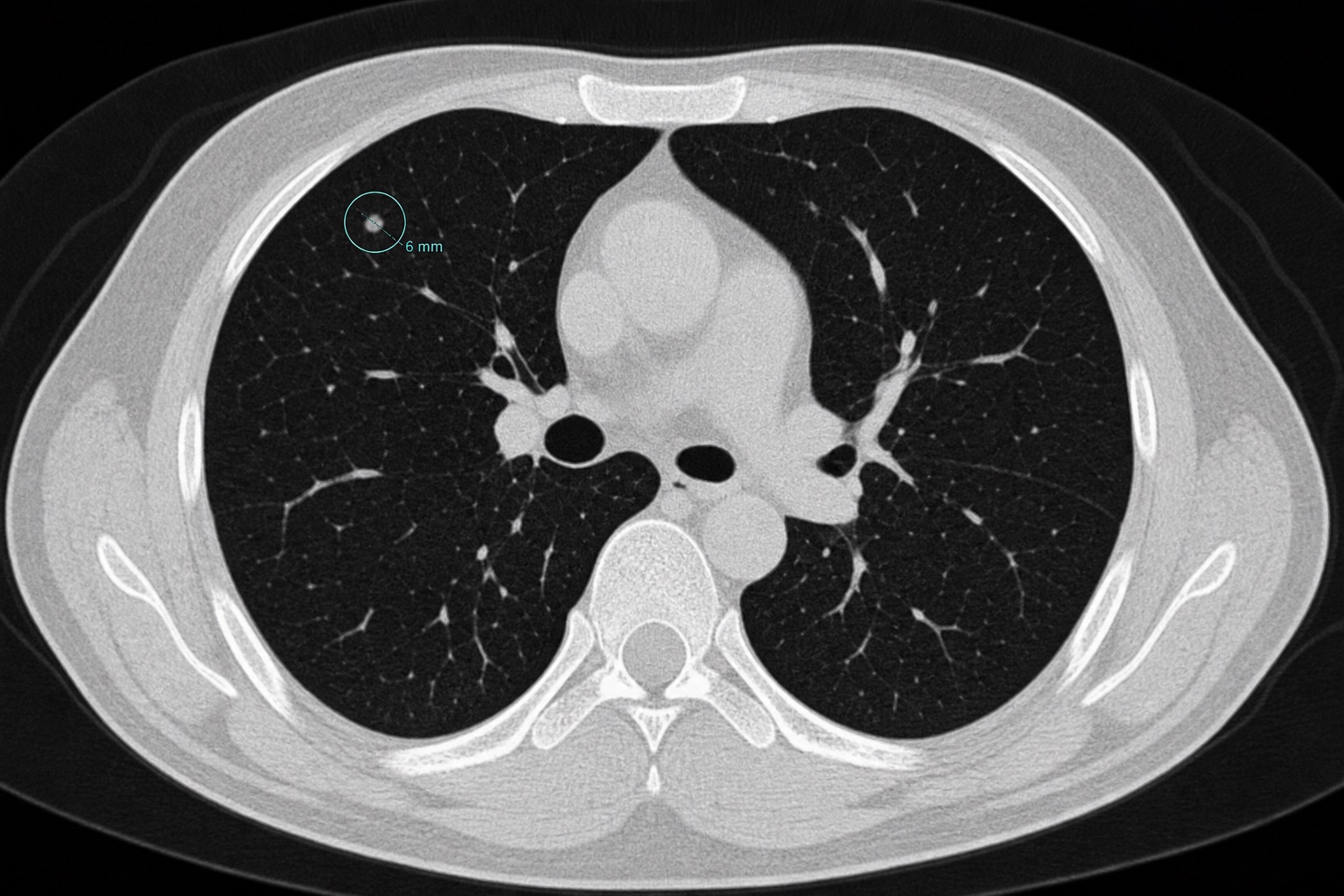
Lung Nodule Size Chart and When to Worry
A clear lung nodule size chart for CT findings: <4 mm, 4-6 mm, 6-8 mm, >8 mm, growth criteria, cancer risk features, and Fleischner follow-up.
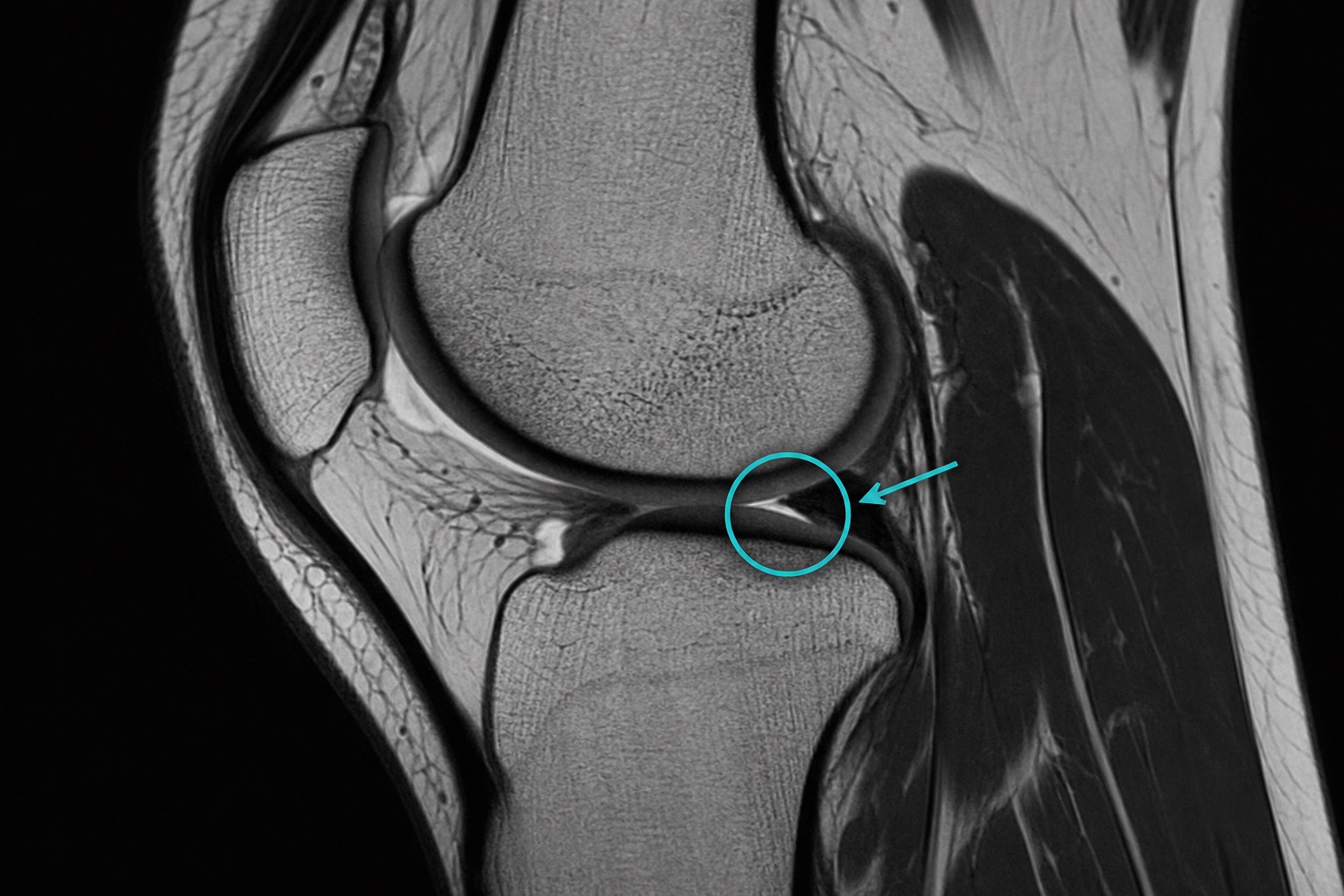
Torn Meniscus on MRI: How to Tell
Learn how a torn meniscus on MRI appears: meniscus tear signal, posterior horn tears, bucket-handle tear signs, grading, and treatment clues.
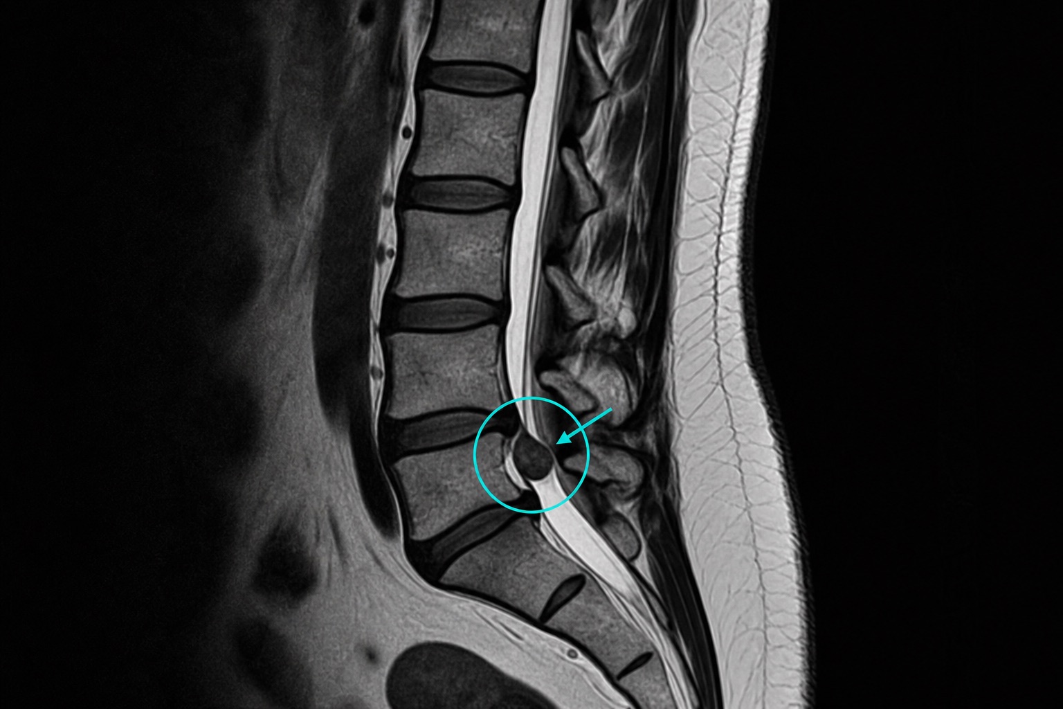
Herniated Disc on MRI: Reading Tips
Understand a herniated disc on MRI: disc bulge vs protrusion vs extrusion, lumbar MRI levels, nerve root compression, spinal stenosis, and red flags.
CT Read Blog
Understand medical images before your next appointment
The CT Read Blog is organized around high-intent search topics: CT scan interpretation, MRI interpretation, chest X-ray interpretation, lung nodule follow-up, brain MRI white matter findings, knee MRI meniscus tears, and lumbar spine MRI disc herniation. These medical imaging interpretation guides explain what the finding looks like, what body problems it can suggest, when it is urgent, and which follow-up test is usually considered.
CT scan interpretation
Learn CT scan orientation, Hounsfield units, contrast phases, lung nodule size thresholds, and red-flag findings that should be discussed with a doctor.
MRI interpretation
Learn MRI sequences like T1, T2, FLAIR, DWI and contrast-enhanced MRI, including how brain MRI, knee MRI and lumbar MRI findings are described.
X-ray interpretation
Use systematic X-ray reading methods for chest X-ray, bone X-ray and emergency findings such as pneumothorax, fracture, effusion, and consolidation.
DICOM and imaging tools
Open DICOM images, inspect metadata, convert DICOM to JPG or video, and prepare imaging files for second opinions, teaching, research, or AI analysis.
How to use these radiology guides
Start with the article that matches the exact phrase in your radiology report. If your report says “white matter hyperintensities,” read the brain MRI white spots guide. If it says “solid lung nodule 6 mm,” use the lung nodule size chart. If it says “posterior horn meniscus tear,” read the torn meniscus MRI guide. Then compare the article with your official report and bring specific questions to your radiologist, primary doctor, pulmonologist, neurologist, orthopedic surgeon, or spine specialist.