
ما هي البقع البيضاء في التصوير بالرنين المغناطيسي للدماغ؟
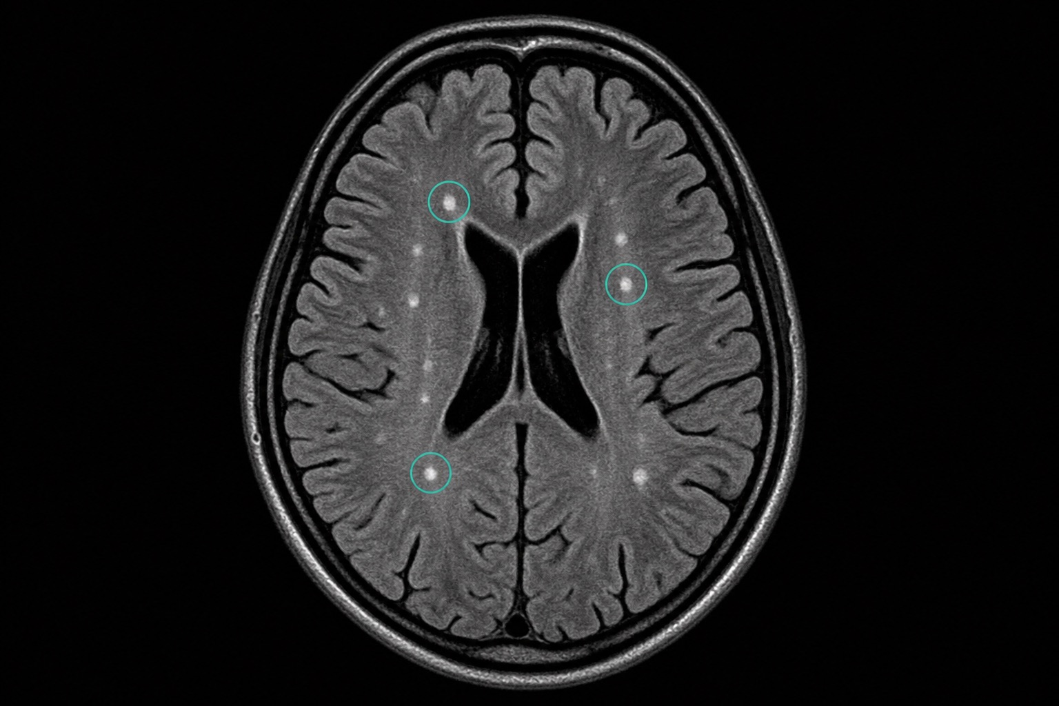
قد يكون رؤية مصطلحات مثل "فرط إشارات T2/FLAIR" أو "فرط إشارات المادة البيضاء" في تقرير التصوير بالرنين المغناطيسي للدماغ أمرًا مخيفًا. تظهر هذه على شكل بقع بيضاء في الصورة وتشير إلى تغيرات طفيفة في محتوى الماء في أنسجة الدماغ.
الأسباب الشائعة
- مرض الأوعية الدموية الدماغية الصغيرة (CSVD): السبب الأكثر شيوعًا، ويرتبط بالشيخوخة وارتفاع ضغط الدم وارتفاع الكوليسترول.
- الصداع النصفي: غالبًا ما يعاني مرضى الصداع النصفي المزمن من بقع بيضاء حميدة في التصوير بالرنين المغناطيسي للدماغ.
- التصلب المتعدد (MS): مرض مزيل للميالين حيث تتجمع البقع البيضاء في مسارات محددة للمادة البيضاء.
- التصلب الدبقي: نسيج ندبي يتبقى من الإصابات الدقيقة السابقة في الدماغ.
Frequently asked questions
مقالات ذات صلة
كيفية قراءة التصوير المقطعي المحوسب: دليل المبتدئين
تعلم كيفية قراءة التصوير المقطعي المحوسب خطوة بخطوة: الشرائح المحورية، والمناظر الإكليلية والساجيتالية، ووحدات هاونسفيلد، وإعدادات نافذة التصوير المقطعي المحوسب، والتشريح الرئيسي، والعلامات الحمراء العاجلة.
كيفية قراءة التصوير بالرنين المغناطيسي
تعلم كيفية قراءة التصوير بالرنين المغناطيسي بلغة مبسطة: T1 مقابل T2، FLAIR، DWI/ADC، تعزيز التباين، مستويات الصورة، القطع الأثرية، ومصطلحات التقرير.
كيفية قراءة الأشعة السينية للصدر
تعلم كيفية قراءة الأشعة السينية للصدر باستخدام طريقة ABCDE المنهجية: جودة الصورة، مجرى الهواء، الرئتين، حجم القلب، الحجاب الحاجز، التشوهات الشائعة، والعلامات الحمراء.