
Introduction
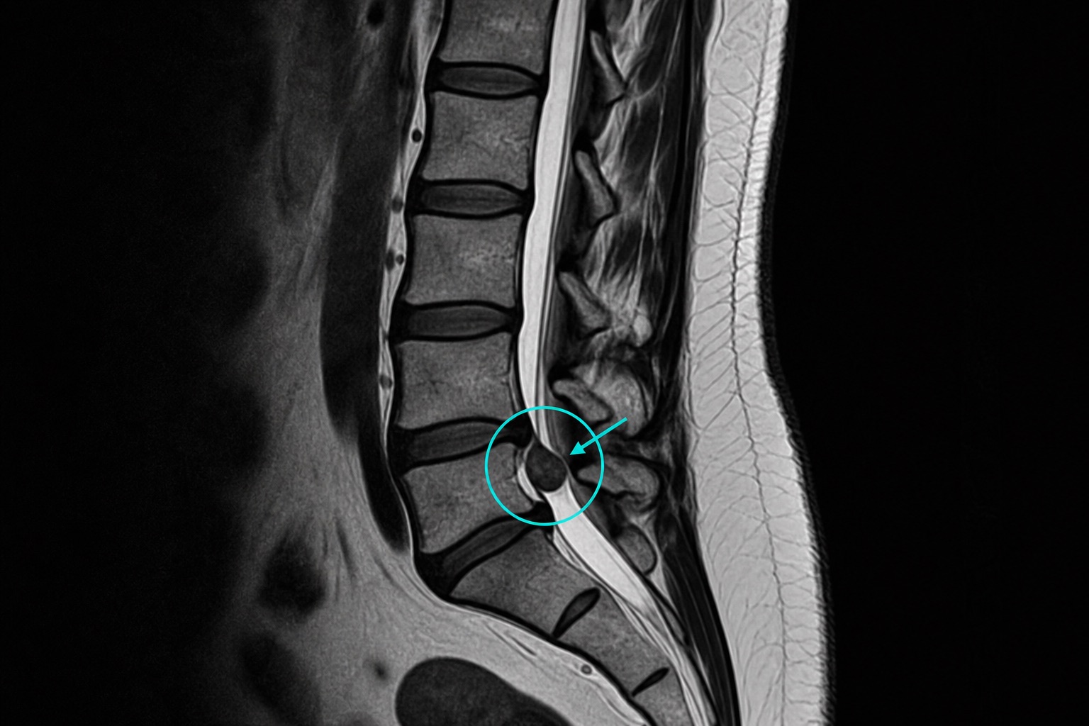
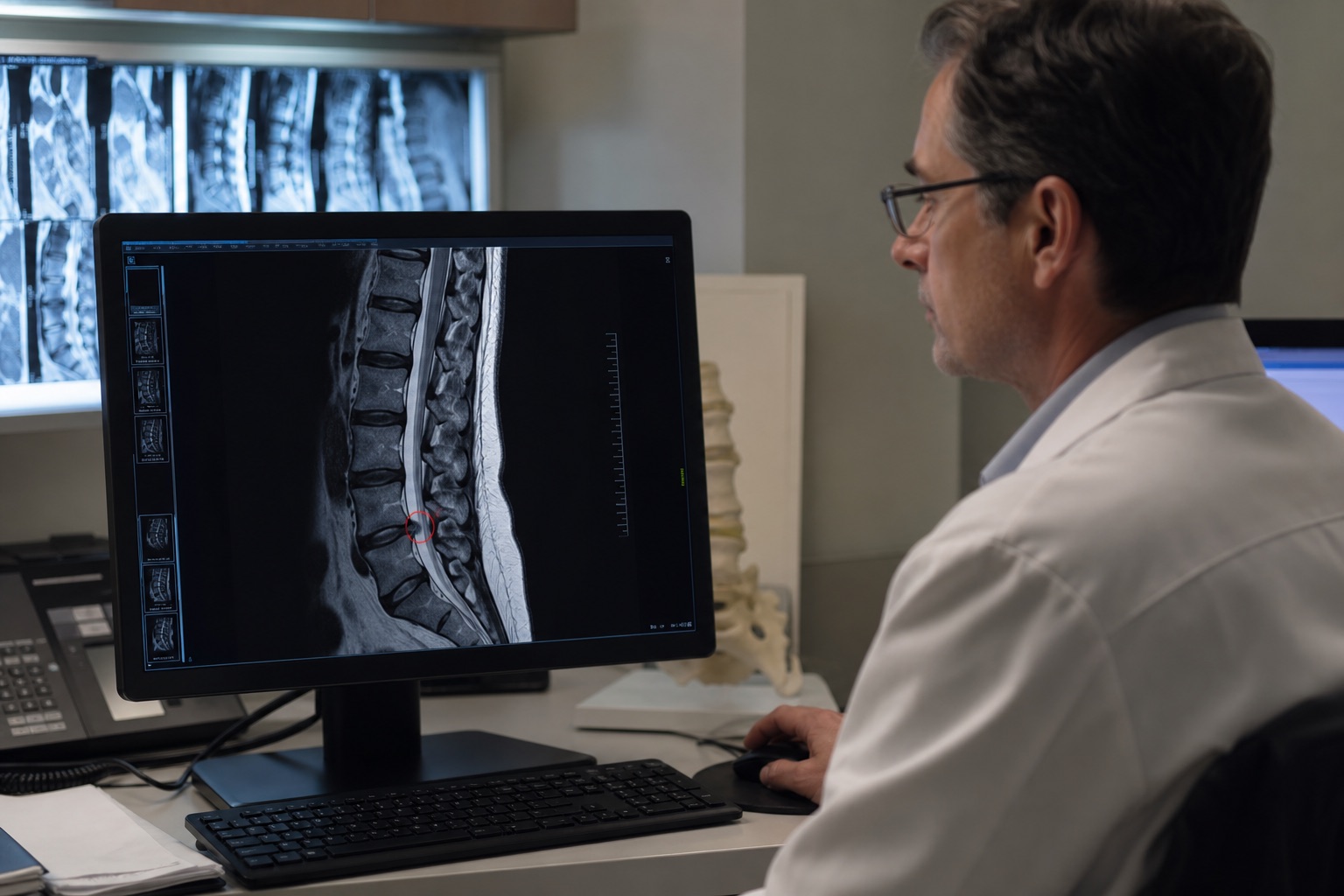
A herniated disc occurs when the soft inner core of a spinal disc pushes through the outer ring, compressing spinal nerves and causing sciatica. MRI is the best imaging modality to evaluate disc herniations.
Disc Pathology Grading on MRI
- Bulging: The disc extends symmetrically beyond the vertebral border. Typically mild.
- Protrusion: A focal projection of disc material, but the base remains wider than the dome.
- Extrusion: The projected material narrow at its base, compressing nerves severely. Fragments may sequester (separate from the disc).
Frequently asked questions
Related articles
How to Read a CT Scan: A Beginner's Guide
Learn how to read a CT scan step by step: axial slices, coronal and sagittal views, Hounsfield units, CT window settings, key anatomy, and urgent red flags.
How to Read an MRI Scan
Learn how to read an MRI scan in plain language: T1 vs T2, FLAIR, DWI/ADC, contrast enhancement, image planes, artifacts, and report terminology.
How to Read a Chest X-Ray
Learn how to read a chest X-ray using a systematic ABCDE method: image quality, airway, lungs, heart size, diaphragm, common abnormalities, and red flags.