

Introduction
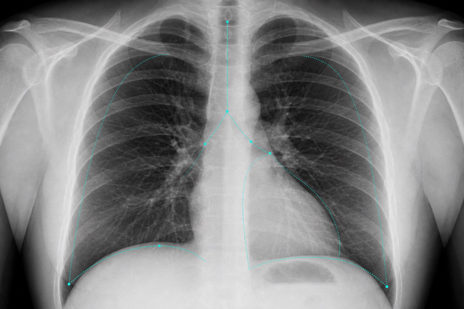
A chest X-ray is one of the most common medical imaging exams. Whether for a routine check-up or due to cough, fever, or chest pain, doctors frequently order a chest film. This guide systemizes how to read a chest X-ray using the ABCDE method.
The ABCDE Systematic Approach
- A (Airway): Check if the trachea is central. Deviations suggest pressure imbalances (e.g., tension pneumothorax).
- B (Breathing): Observe the lungs. Air-filled lungs appear black. White patches suggest fluid, consolidation (pneumonia), or masses.
- C (Circulation): Assess the heart. The heart size should not exceed 50% of the chest diameter.
- D (Diaphragm): Diaphragms should be dome-shaped. Check for free air beneath them and blunt costophrenic angles (indicating effusion).
- E (Everything else): Check ribs, clavicles, and soft tissues for fractures or abnormal air.
Frequently asked questions
Useful tools
Related articles
How to Read a CT Scan: A Beginner's Guide
Learn how to read a CT scan step by step: axial slices, coronal and sagittal views, Hounsfield units, CT window settings, key anatomy, and urgent red flags.
How to Read an MRI Scan
Learn how to read an MRI scan in plain language: T1 vs T2, FLAIR, DWI/ADC, contrast enhancement, image planes, artifacts, and report terminology.
White Spots on Brain MRI: What They Mean
Understand white spots on brain MRI: T2/FLAIR white matter hyperintensities, migraine spots, small vessel disease, MS patterns, risk factors, and follow-up.