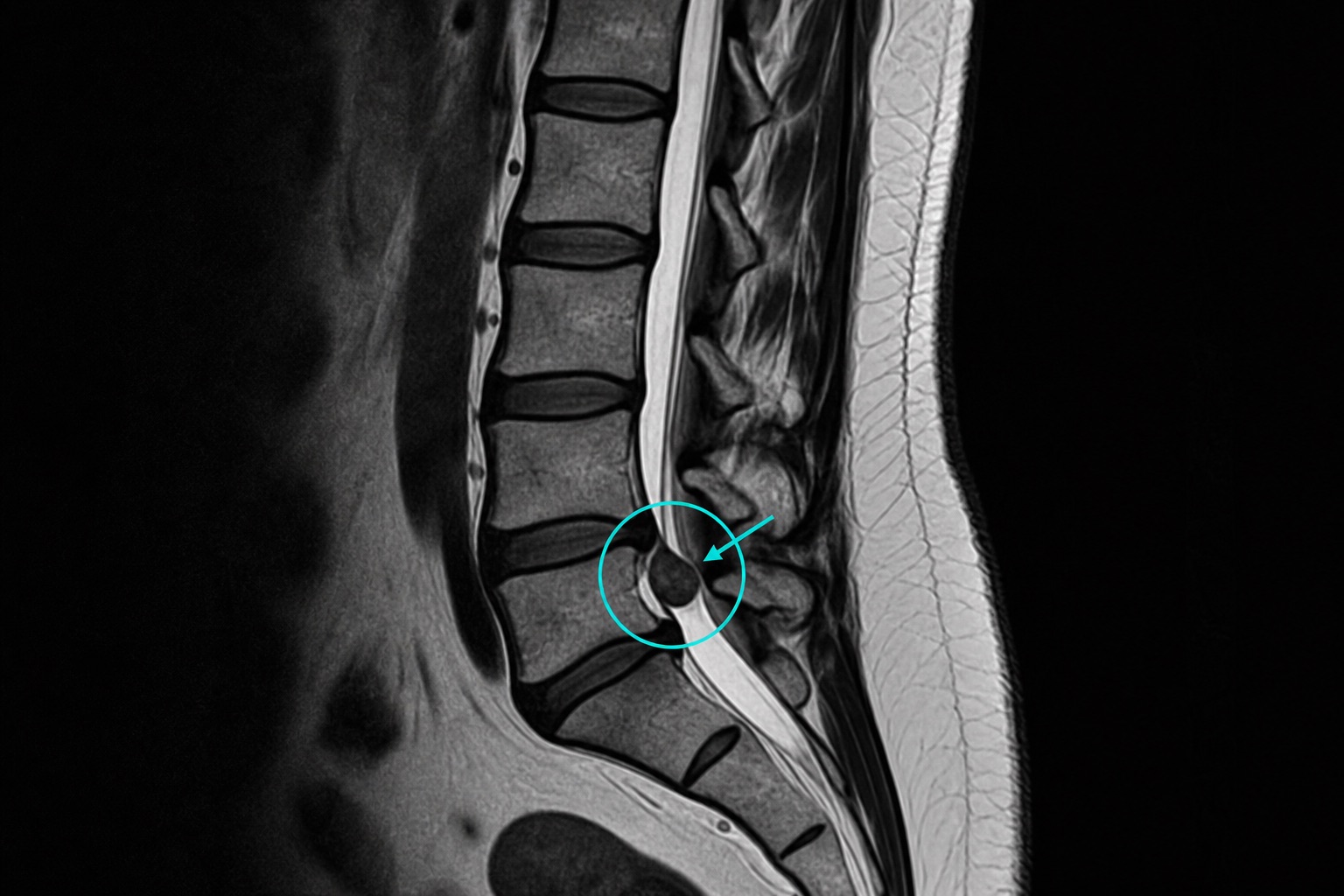
Introduction
A herniated disc happens when the soft center (nucleus pulposus) of a spinal disc pushes through its tougher outer ring (annulus fibrosus). MRI is the best test for evaluating the spine and nerve roots. This guide will help you decode the most common terms used in lumbar MRI reports — bulge, protrusion, extrusion, sequestration, and how they relate to nerve compression.
If your report mentions a herniated disc on MRI, the most important details are the level, the shape of the disc material, and whether a nerve root is actually compressed. A herniated disc on MRI may sound alarming, but disc bulges, protrusions, and even small extrusions can appear in people without pain. The MRI finding becomes clinically important when it matches leg pain, numbness, weakness, or reflex changes in the same nerve distribution.
Key takeaways about a herniated disc on MRI
- A herniated disc on MRI is described by shape. Bulge, protrusion, extrusion, and sequestration each mean something different.
- A herniated disc on MRI is described by location. Central, paracentral, foraminal, and extraforaminal disc herniations affect different nerve roots.
- A herniated disc on MRI needs clinical correlation. Imaging findings only matter if they explain the patient's symptoms and exam.
- A herniated disc on MRI can improve. Many extrusions and sequestered fragments shrink over months with conservative care.
1. Disc terminology in plain English
- Bulge: smooth, generalized extension of the disc beyond the vertebral body, involving more than 25% of the disc circumference. Often a normal aging finding.
- Protrusion: focal extension where the base is wider than the apex. The annulus is intact.
- Extrusion: focal extension where the apex is wider than the base, suggesting a tear in the annulus.
- Sequestration: a free fragment of disc material has separated from the parent disc and migrated.
2. Why location matters
Reports describe disc location relative to the spinal canal: central, paracentral, subarticular, foraminal, or extraforaminal. The location helps predict which nerve root is compressed.
- Central: may compress the cauda equina (medical emergency if severe).
- Paracentral / subarticular: typically affects the traversing nerve root (e.g., L5 root at L4-L5).
- Foraminal: typically affects the exiting nerve root (e.g., L4 root at L4-L5).
3. Other key findings on lumbar MRI
- Spinal canal stenosis: narrowing of the central canal, often by combined disc and ligament hypertrophy.
- Foraminal stenosis: narrowing of the nerve root exit, often causing radiculopathy.
- Modic changes: signal changes in vertebral end plates from disc degeneration.
- Facet joint arthrosis: degenerative joint changes that can contribute to back pain.
4. Red flag findings
- Cauda equina syndrome: severe central canal compression with saddle anesthesia, urinary retention, or new bowel/bladder dysfunction. Surgical emergency.
- Progressive motor weakness in a nerve root distribution.
- Suspected infection or tumor (replacement of marrow signal, paraspinal mass).
5. Conservative vs surgical management
Most lumbar disc herniations improve with conservative care: rest, NSAIDs, physical therapy, and time. Surgery (microdiscectomy) is considered for persistent radicular pain > 6 weeks despite conservative care, progressive neurological deficit, or red flag findings. The MRI is just one input — clinical correlation is essential.
For an AI-powered explanation of your lumbar MRI report, try our spine MRI interpretation tool.
When reading about a herniated disc on MRI, look for phrases like “contacting,” “displacing,” “compressing,” or “impinging” the nerve root. These words tell you whether the herniated disc is merely present or is likely causing radiculopathy.
Frequently asked questions
Related articles
How to Read a CT Scan: A Beginner’s Guide
Learn how to read a CT scan step by step: axial slices, coronal and sagittal views, Hounsfield units, CT window settings, key anatomy, and urgent red flags.
How to Read an MRI Scan
Learn how to read an MRI scan in plain language: T1 vs T2, FLAIR, DWI/ADC, contrast enhancement, image planes, artifacts, and report terminology.
How to Read a Chest X-Ray
Learn how to read a chest X-ray using a systematic ABCDE method: image quality, airway, lungs, heart size, diaphragm, common abnormalities, and red flags.


