
Introduction
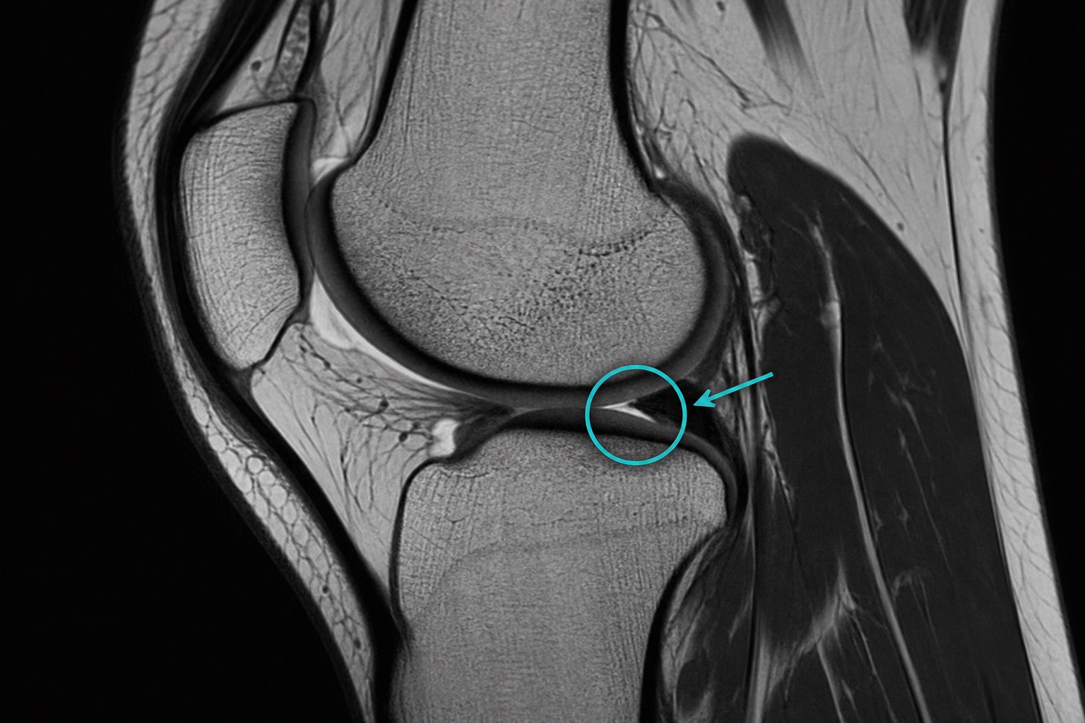
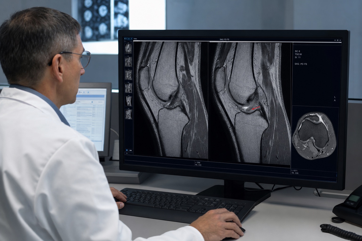
The meniscus is a C-shaped cartilage pad in the knee joint. Meniscus tears are common orthopedic injuries diagnosed with T2 or PD weighted MRI sequences.
MRI Visual Clues for Meniscus Tears
- Abnormal High Signal Touching the Joint Surface: The golden standard for a tear on MRI is a white line inside the black meniscus triangle that extends to the upper or lower boundary.
- Morphological Distortion: The meniscus appears frayed, displaced, or significantly smaller than expected.
Frequently asked questions
Related articles
How to Read a CT Scan: A Beginner's Guide
Learn how to read a CT scan step by step: axial slices, coronal and sagittal views, Hounsfield units, CT window settings, key anatomy, and urgent red flags.
How to Read an MRI Scan
Learn how to read an MRI scan in plain language: T1 vs T2, FLAIR, DWI/ADC, contrast enhancement, image planes, artifacts, and report terminology.
How to Read a Chest X-Ray
Learn how to read a chest X-ray using a systematic ABCDE method: image quality, airway, lungs, heart size, diaphragm, common abnormalities, and red flags.