
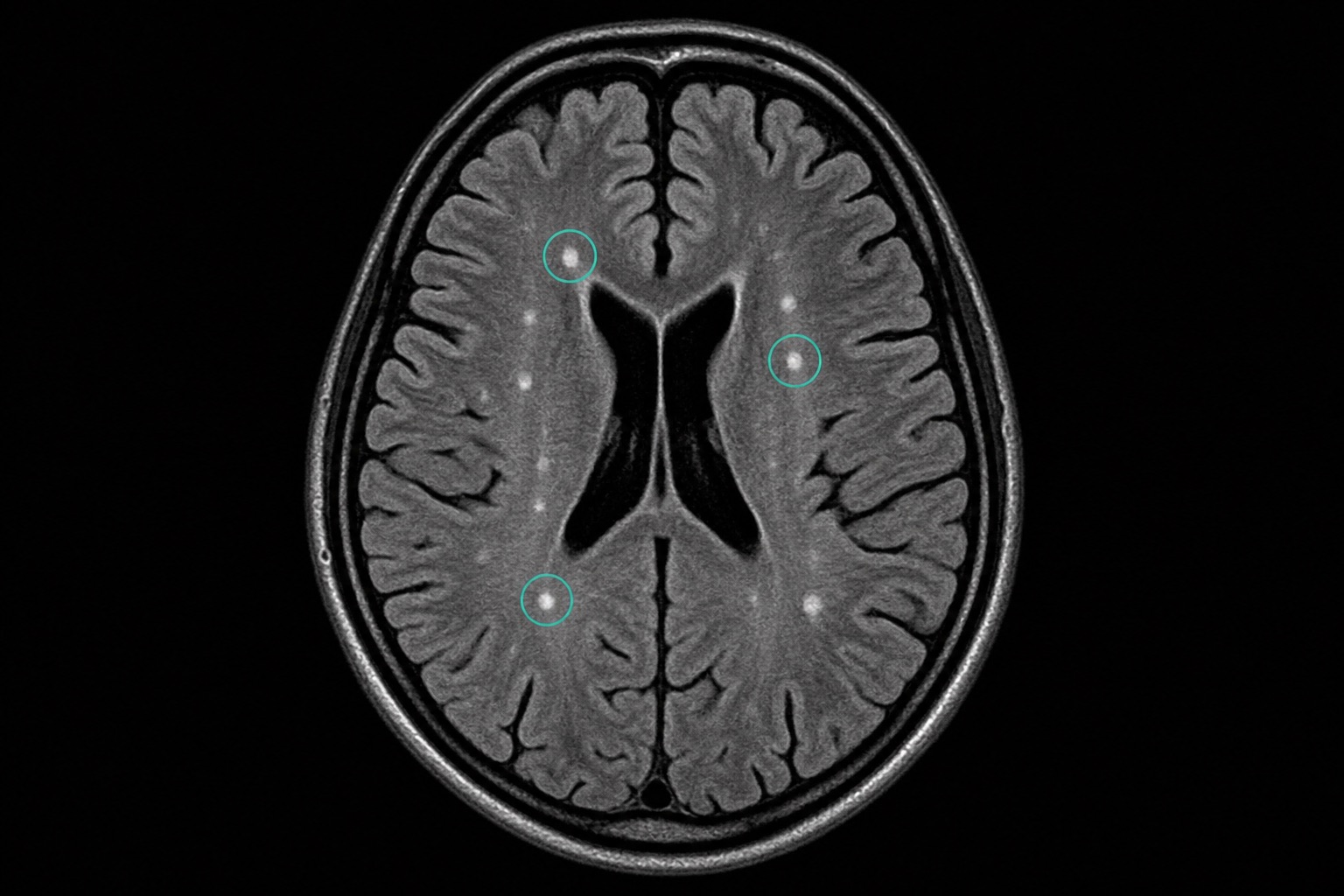

What are White Spots on Brain MRI?
Seeing terms like "T2/FLAIR hyperintensities" or "white matter hyperintensities" in a brain MRI report can be scary. These appear as white spots on the image and indicate subtle water content changes in the brain tissue.
Common Causes
- Cerebral Small Vessel Disease (CSVD): The most common cause, linked to aging, hypertension, and high cholesterol.
- Migraines: Chronic migraine sufferers often have benign white spots on their brain MRI.
- Multiple Sclerosis (MS): A demyelinating disease where white spots cluster in specific white matter pathways.
- Gliosis: Scar tissue left behind from previous micro-injuries in the brain.
Frequently asked questions
Related articles
How to Read a CT Scan: A Beginner's Guide
Learn how to read a CT scan step by step: axial slices, coronal and sagittal views, Hounsfield units, CT window settings, key anatomy, and urgent red flags.
How to Read an MRI Scan
Learn how to read an MRI scan in plain language: T1 vs T2, FLAIR, DWI/ADC, contrast enhancement, image planes, artifacts, and report terminology.
How to Read a Chest X-Ray
Learn how to read a chest X-ray using a systematic ABCDE method: image quality, airway, lungs, heart size, diaphragm, common abnormalities, and red flags.