Introduction
Une radiographie pulmonaire est l'un des examens d'imagerie médicale les plus courants. Que ce soit pour un bilan de routine ou en raison de toux, de fièvre ou de douleurs thoraciques, les médecins prescrivent fréquemment une radiographie pulmonaire. Ce guide systématise la lecture d'une radiographie pulmonaire en utilisant la méthode ABCDE.
L'approche systématique ABCDE
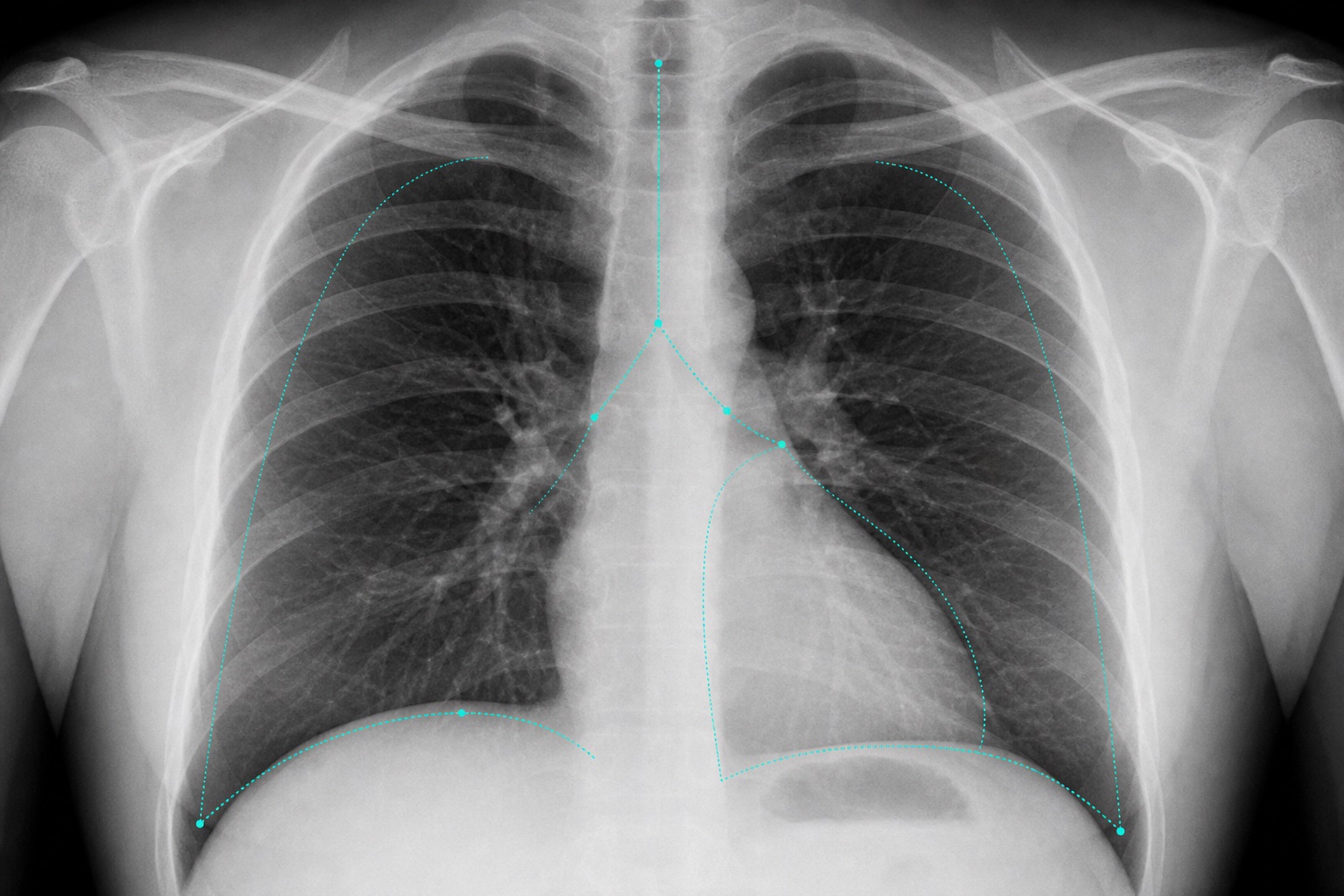
- A (Voies aériennes) : Vérifiez si la trachée est centrale. Les déviations suggèrent des déséquilibres de pression (par exemple, un pneumothorax sous tension).
- B (Respiration) : Observez les poumons. Les poumons remplis d'air apparaissent noirs. Les taches blanches suggèrent du liquide, une consolidation (pneumonie) ou des masses.
- C (Circulation) : Évaluez le cœur. La taille du cœur ne doit pas dépasser 50 % du diamètre thoracique.
- D (Diaphragme) : Les diaphragmes doivent être en forme de dôme. Vérifiez la présence d'air libre sous ceux-ci et l'émoussement des angles costo-phréniques (indiquant un épanchement).
- E (Tout le reste) : Vérifiez les côtes, les clavicules et les tissus mous pour des fractures ou de l'air anormal.
Frequently asked questions
Outils utiles
Articles connexes
Comment lire un scanner CT : un guide pour débutants
Apprenez à lire un scanner CT étape par étape : coupes axiales, vues coronales et sagittales, unités Hounsfield, réglages de fenêtre CT, anatomie clé et signes d'alerte urgents.
Comment lire une IRM
Apprenez à lire une IRM en langage clair : T1 vs T2, FLAIR, DWI/ADC, rehaussement de contraste, plans d'image, artefacts et terminologie des rapports.
Taches blanches sur l'IRM cérébrale : ce qu'elles signifient
Comprendre les taches blanches sur l'IRM cérébrale : hyperintensités de la substance blanche T2/FLAIR, taches de migraine, maladie des petits vaisseaux, schémas de la SEP, facteurs de risque et suivi.