
Introduzione
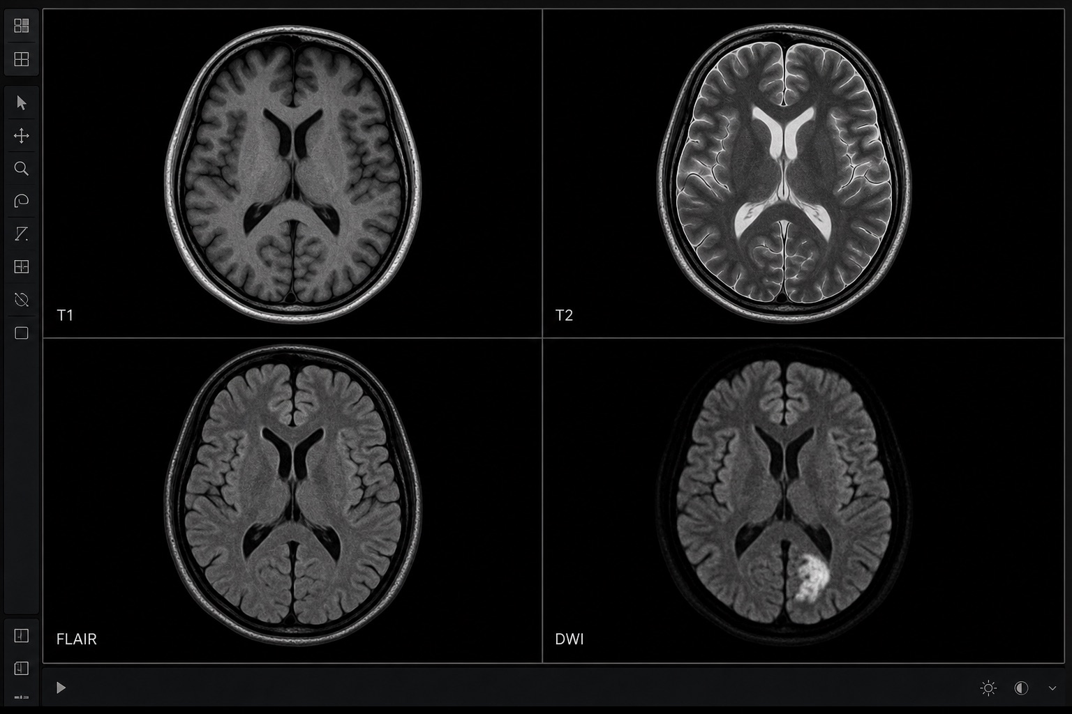
La risonanza magnetica (RM) crea immagini dettagliate utilizzando potenti magneti e onde radio, senza radiazioni. Il compromesso è la complessità: ogni studio RM contiene più sequenze (T1, T2, FLAIR, DWI, T1 con contrasto...) che evidenziano tessuti diversi. Sapere cosa mostra ogni sequenza fa un'enorme differenza nella comprensione del referto.
Se vuoi imparare come leggere una risonanza magnetica, inizia pensando in sequenze anziché in colori. Una TAC chiede principalmente "quanto è densa questa struttura?"; la RM chiede "quale proprietà del tessuto viene evidenziata da questa sequenza?". Ecco perché l'interpretazione della RM dipende dalla ripetizione della stessa domanda attraverso le immagini T1, T2, FLAIR, DWI/ADC, STIR e con contrasto.
Punti chiave prima di leggere una risonanza magnetica
- L'interpretazione della RM è basata sulle sequenze. Le immagini T1, T2, FLAIR, DWI, ADC, STIR e post-contrasto rispondono ciascuna a una domanda diversa.
- Le anomalie della RM dovrebbero essere controllate su più piani. Le viste assiali, sagittali e coronali aiutano a separare le vere lesioni dagli artefatti.
- Il segnale RM non è una diagnosi di per sé. Un'iperintensità T2, una diffusione ristretta o un pattern di enhancement con contrasto necessitano di contesto clinico.
- I referti RM riassumono il reperto importante nell'Impressione. Leggi prima il corpo del referto, quindi usa l'Impressione per capire cosa conta di più.
1. T1 vs T2 — la guida rapida
Due sequenze di base coprono la maggior parte di ciò che i pazienti vedono nella loro risonanza magnetica:
- Ponderata in T1: il fluido è scuro (liquor, urina), il grasso è brillante. Ottima per l'anatomia.
- Ponderata in T2: il fluido è brillante, il grasso è scuro. Eccellente per individuare edema, infiammazione e la maggior parte dei tumori (che contengono acqua extra).
Il trucco: se una struttura è brillante in T2 e scura in T1, è probabilmente ricca di acqua (cisti, edema, liquor). Se è brillante in T1 e scura in T2, pensa a grasso o prodotti ematici cronici.
2. Altre sequenze comuni
- FLAIR — come T2 ma sopprime il liquor. Le lesioni della sostanza bianca nella SM risaltano.
- DWI/ADC — imaging pesato in diffusione. Rileva ictus acuto e ascessi mostrando un movimento dell'acqua ristretto.
- STIR — sequenza con soppressione del grasso; ottima per l'edema del midollo osseo.
- T1 + Contrasto (gadolinio) — evidenzia tumori, infiammazioni e barriera emato-encefalica alterata.
3. Leggere su 3 piani — assiale, sagittale, coronale
Proprio come la TAC, la RM viene ricostruita in tre piani ortogonali. Le lesioni di solito sembrano reali quando è possibile identificarle su più di un piano. Un reperto che appare solo su una singola fetta è spesso un artefatto.
4. Cosa significa "enhancement con contrasto"?
Il contrasto di gadolinio si accumula dove i vasi sanguigni sono anomali — tipicamente tumori, infezioni e demielinizzazione attiva. Il referto potrebbe usare frasi come "enhancement ad anello" (spesso ascessi o tumori) o "enhancement avido" (tumori vascolari). La mancanza di enhancement aiuta a escludere determinate patologie.
5. Un approccio sistematico
- Confermare nome del paziente, parte del corpo e data dello studio.
- Identificare le sequenze elencate nel referto (spesso abbreviate: T1, T2, FLAIR, DWI, T1+Gd).
- Confrontare la lesione sospetta tra sequenze e piani.
- Leggere la sezione "Impressione" per ultima — riassume il significato clinico.
Passi successivi
Per pagine specifiche sulla RM, consulta le nostre guide sulla RM cerebrale, RM del ginocchio o RM lombare — oppure carica la tua scansione direttamente sul nostro strumento di analisi RM AI.
Il modo più sicuro per esercitarsi a leggere una risonanza magnetica è confrontare una sequenza alla volta con il referto radiologico. Se un reperto appare in T2 ma non in FLAIR, o brillante in DWI ma non scuro in ADC, il significato cambia. Questo è il motivo per cui la lettura della RM è un processo di riconoscimento di pattern piuttosto che un giudizio su una singola immagine.
Frequently asked questions
Strumenti utili
Articoli correlati
Come leggere una TC: una guida per principianti
Impara a leggere una TC passo dopo passo: sezioni assiali, viste coronali e sagittali, unità Hounsfield, impostazioni della finestra TC, anatomia chiave e segnali di allarme urgenti.
Come leggere una radiografia del torace
Impara a leggere una radiografia del torace usando un metodo sistematico ABCDE: qualità dell'immagine, vie aeree, polmoni, dimensioni del cuore, diaframma, anomalie comuni e segnali di allarme.
Macchie bianche sulla risonanza magnetica cerebrale: cosa significano
Comprendi le macchie bianche sulla risonanza magnetica cerebrale: iperintensità della sostanza bianca T2/FLAIR, macchie da emicrania, malattia dei piccoli vasi, pattern di SM, fattori di rischio e follow-up.