
Pendahuluan
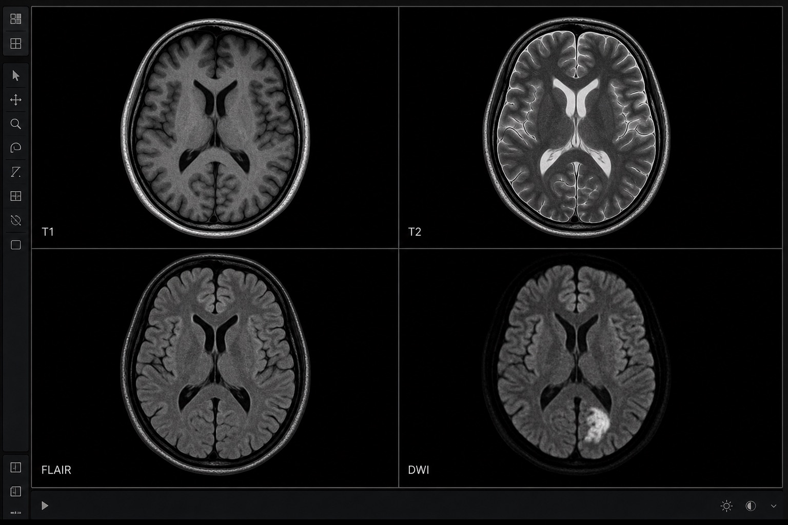
MRI (magnetic resonance imaging) menghasilkan gambaran detail menggunakan magnet kuat dan gelombang radio — tanpa radiasi. Kekurangannya adalah kompleksitas: setiap studi MRI berisi beberapa sekuens (T1, T2, FLAIR, DWI, T1 dengan kontras...) yang menyoroti jaringan yang berbeda. Mengetahui apa yang ditunjukkan oleh setiap sekuens membuat perbedaan besar dalam memahami laporan Anda.
Jika Anda ingin belajar cara membaca MRI, mulailah dengan berpikir dalam sekuens, bukan warna. CT scan sebagian besar bertanya “seberapa padat struktur ini?”; MRI bertanya “properti jaringan mana yang disorot oleh sekuens ini?” Itulah mengapa interpretasi MRI bergantung pada pengulangan pertanyaan yang sama di seluruh gambar T1, T2, FLAIR, DWI/ADC, STIR, dan kontras.
Poin-poin penting sebelum membaca MRI
- Interpretasi MRI berbasis sekuens. Gambar T1, T2, FLAIR, DWI, ADC, STIR, dan pasca-kontras masing-masing menjawab pertanyaan yang berbeda.
- Kelainan MRI harus diperiksa dalam lebih dari satu bidang. Pandangan aksial, sagital, dan koronal membantu memisahkan lesi sejati dari artefak.
- Sinyal MRI bukanlah diagnosis itu sendiri. Pola hiperintensitas T2, difusi terbatas, atau peningkatan kontras memerlukan konteks klinis.
- Laporan MRI merangkum temuan penting dalam Kesimpulan. Baca isi laporan terlebih dahulu, lalu gunakan Kesimpulan untuk memahami apa yang paling penting.
1. T1 vs T2 — panduan singkat
Dua sekuens dasar mencakup sebagian besar apa yang pasien lihat pada MRI mereka:
- T1-weighted: cairan gelap (CSF, urin), lemak terang. Baik untuk anatomi.
- T2-weighted: cairan terang, lemak redup. Sangat baik untuk melihat edema, peradangan, dan sebagian besar tumor (yang mengandung air ekstra).
Triknya: jika suatu struktur terang pada T2 dan gelap pada T1, kemungkinan besar kaya air (kista, edema, CSF). Jika terang pada T1 dan gelap pada T2, pikirkan lemak atau produk darah kronis.
2. Sekuens umum lainnya
- FLAIR — seperti T2 tetapi menekan CSF. Lesi materi putih pada MS muncul.
- DWI/ADC — pencitraan tertimbang difusi. Mendeteksi stroke akut dan abses dengan menunjukkan gerakan air yang terbatas.
- STIR — sekuens penekan lemak; bagus untuk edema sumsum tulang.
- T1 + Kontras (gadolinium) — menyoroti tumor, peradangan, dan gangguan sawar darah-otak.
3. Baca dalam 3 bidang — aksial, sagital, koronal
Sama seperti CT, MRI direkonstruksi dalam tiga bidang ortogonal. Lesi biasanya terlihat nyata ketika Anda dapat mengidentifikasinya pada lebih dari satu bidang. Temuan yang hanya muncul pada satu irisan seringkali merupakan artefak.
4. Apa arti “meningkat dengan kontras”?
Kontras gadolinium terakumulasi di mana pembuluh darah abnormal — biasanya tumor, infeksi, dan demielinasi aktif. Laporan Anda mungkin menggunakan frasa seperti “peningkatan cincin” (seringkali abses atau tumor) atau “peningkatan avid” (tumor vaskular). Kurangnya peningkatan membantu menyingkirkan patologi tertentu.
5. Pendekatan sistematis
- Konfirmasi nama pasien, bagian tubuh, dan tanggal studi.
- Identifikasi sekuens yang tercantum dalam laporan (sering disingkat: T1, T2, FLAIR, DWI, T1+Gd).
- Bandingkan lesi yang dicurigai di seluruh sekuens dan bidang.
- Baca bagian “Kesimpulan” terakhir — ini merangkum signifikansi klinis.
Langkah selanjutnya
Untuk sub-halaman MRI tertentu, lihat panduan MRI otak, MRI lutut, atau MRI lumbal kami — atau unggah pindaian Anda langsung ke alat analisis MRI AI kami.
Cara teraman untuk berlatih cara membaca MRI adalah dengan membandingkan satu sekuens pada satu waktu dengan laporan radiologi. Jika suatu temuan muncul pada T2 tetapi tidak pada FLAIR, atau terang pada DWI tetapi tidak gelap pada ADC, artinya berubah. Inilah mengapa pembacaan MRI adalah proses pencocokan pola daripada penilaian satu gambar.
Frequently asked questions
Alat yang berguna
Artikel terkait
Cara Membaca Pemindaian CT: Panduan Pemula
Pelajari cara membaca pemindaian CT langkah demi langkah: irisan aksial, tampilan koronal dan sagital, unit Hounsfield, pengaturan jendela CT, anatomi kunci, dan tanda bahaya mendesak.
Cara Membaca Rontgen Dada
Pelajari cara membaca rontgen dada menggunakan metode ABCDE sistematis: kualitas gambar, saluran napas, paru-paru, ukuran jantung, diafragma, kelainan umum, dan tanda bahaya.
Bintik Putih pada MRI Otak: Apa Artinya
Pahami bintik putih pada MRI otak: hiperintensitas materi putih T2/FLAIR, bintik migrain, penyakit pembuluh darah kecil, pola MS, faktor risiko, dan tindak lanjut.