AI 부비동 X선 판독
부비동 X선 사진이 무엇을 보여주는지 평이한 언어로 이해하는 데 도움을 받으세요. 부비동, 점막 비후 및 부비동염과 관련된 공기-액체 경계면에 대한 명확한 설명.
수천 명의 사람들이 부비동 X선 결과를 이해하도록 돕습니다.
이 도구는 X선에서 부비동을 식별하고 부비동염 또는 공기-액체 경계면과 같은 일반적인 소견에 대한 간단한 설명을 제공합니다.
부비동 X선 판독이란 무엇인가요?
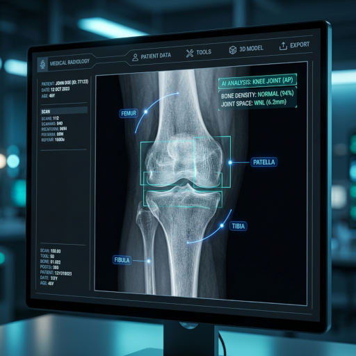
부비동 X선(종종 Waters' view)은 부비동의 선별 영상 검사입니다. CT Read는 염증이나 체액의 징후를 포함하여 상악동, 전두동, 사골동 및 접형동의 모양을 이해하는 데 도움을 줍니다.
상악동
뺨뼈에 위치한 상악동은 부비동염에 가장 흔하게 영향을 받는 부위입니다. 이곳의 점막 비후 또는 공기-액체 경계면은 주요 소견입니다.
전두동
눈썹 위에 위치한 전두동은 전두동염과 함께 불투명화 또는 공기-액체 경계면을 보일 수 있습니다. 발달은 연령에 따라 다릅니다.
사골동 및 접형동
이 더 깊은 부비동은 일반 X선에서 부분적으로 보입니다. 상세한 평가를 위해서는 일반적으로 CT 스캔이 필요하지만, X선으로도 병리를 시사할 수 있습니다.
공기-액체 경계면 및 불투명화
수평 공기-액체 경계면은 분비물 저류를 동반한 급성 부비동염을 강력히 시사합니다. 미만성 불투명화는 만성 부비동 질환 또는 용종을 나타낼 수 있습니다.
부비동 X선 판독을 쉽게
X선 판독 도구는 간단한 언어를 사용하여 기본적인 부비동 X선 소견을 이해하는 데 도움을 줍니다.
부비동 X선에 대한 간단한 설명
부비동 X선이 무엇을 나타내는지 명확하고 전문 용어가 없는 설명을 받으세요.
부비동 해부학에 대한 교육적 초점
부비동 해부학의 기본과 부비동염의 일반적인 소견을 배우세요.
부비동 X선 판독으로 마음의 평화
의사와 상담하기 전에 부비동 X선 결과에 대한 기본적인 이해를 얻어 불안감을 줄이세요.
부비동 X선 판독 서비스 이용 방법
AI 분석 시스템을 통해 부비동 X선 판독 보고서를 받는 네 가지 간단한 단계:
부비동 X선 업로드
부비동 X선 이미지(Waters' view 권장)를 보안 판독 플랫폼에 업로드하세요.
AI 판독 처리
AI 시스템은 부비동을 분석하여 불투명화, 점막 비후 및 공기-액체 경계면을 찾습니다.
상세 판독 생성
시스템은 소견 및 설명이 포함된 이해하기 쉬운 판독 보고서를 생성합니다.
판독 보기 및 공유
결과를 보고 선택적으로 의사 또는 이비인후과 전문의와 보고서를 안전하게 공유하세요.
부비동 X선 이해하기
부비동 X선 이미지를 업로드하여 이해하기 쉬운 설명을 받으세요.
X-ray, CT, MRI, 초음파
부비동 X선으로 무엇을 감지할 수 있나요?
부비동 X선(Waters', Caldwell, 측면 촬영법)은 얼굴의 상악동, 전두동, 사골동 및 접형동의 공기로 채워진 공동을 평가합니다. 상세한 부비동 평가를 위해 CT가 X선을 대체했지만, 일반 X선 사진은 여전히 유용한 선별 정보를 제공합니다.
공기-액체 경계면을 동반한 급성 부비동염
똑바로 선 Waters' view에서 상악동의 수평 공기-액체 경계면은 급성 세균성 부비동염의 전형적인 징후이며, 종종 항생제 치료를 필요로 합니다.
점막 비후 및 만성 부비동염
5mm 이상의 점막 비후 또는 완전한 부비동 불투명화는 만성 염증을 시사하며, 이는 이비인후과 평가 및 내시경 수술 계획을 위한 CT를 필요로 할 수 있습니다.
부비동 용종 및 저류 낭종
상악동의 매끄럽고 돔 모양의 불투명화는 종종 양성 점액 저류 낭종을 나타냅니다. 더 크거나 불규칙한 용종은 내시경 검사를 필요로 할 수 있습니다.
안면 골절(광대뼈-상악, 코, 안와)
Waters' view는 안면 외상에 대한 표준 선별 촬영법입니다. 이는 안와 바닥 파열("눈물 방울 징후"), 광대뼈 아치 골절 및 코뼈 변위를 보여줍니다.
이물질 및 치과 병리
부비동 또는 비강 내 방사선 불투과성 이물질, 상악동으로 전위된 치아 뿌리, 치성 낭종 모두 식별할 수 있습니다.
원인 불명의 부비동 불투명화
골 파괴를 동반한 한쪽 부비동의 완전한 불투명화는 비강 부비동 종양 또는 침습성 진균성 부비동염을 시사하며, 긴급한 CT/MRI 추적 관찰이 필요합니다.
부비동 X선은 언제 지시되나요?
CT는 이제 대부분의 부비동 문제에 대한 선택 영상 검사이지만, CT를 사용할 수 없을 때 X선은 선별 도구로 여전히 유용합니다.
- 1
급성 안면 통증, 발열 및 화농성 비루
급성 세균성 부비동염이 의심되고 CT를 즉시 사용할 수 없을 때, Waters' view는 공기-액체 경계면을 확인하고 항생제 치료를 뒷받침할 수 있습니다.
- 2
중안면 골절 가능성이 있는 안면 외상
Waters', Caldwell 및 측면 촬영법은 안와 바닥 "폭발" 골절, 광대뼈 아치 골절 및 코 골절을 선별합니다.
- 3
치료에 반응하지 않는 만성 부비동 증상
12주 이상 지속되는 코막힘, 두통 또는 후비루는 영상 검사를 필요로 할 수 있지만, CT가 선호됩니다.
- 4
치아-부비동 교통 의심
발치 또는 신경 치료 후 X선은 구강-상악동 누공 또는 상악동으로 전위된 치아 파편을 감지할 수 있습니다.
- 5
코 또는 부비동 내 이물질 의심
특히 어린이의 경우, 일반 X선은 긴급 제거가 필요한 방사선 불투과성 이물질(단추형 배터리, 자석, 금속)을 감지합니다.
부비동 X선 vs CT vs MRI vs 내시경
부비동 CT는 대부분의 상황에서 현대 표준이 되었지만, X선은 여전히 유용한 선별 도구입니다.
| 영상 촬영 방식 | 가장 잘 보여주는 것 | 한계 | 비용 및 접근성 |
|---|---|---|---|
| 부비동 X선 | 공기-액체 경계면, 불투명화 선별, 이물질, 기본적인 안면 외상 | 대부분의 사골동/접형동 병리를 놓침; 골-구멍 복합체를 상세히 볼 수 없음 | 저렴한 비용, 빠름 |
| 부비동 CT (저선량) | 모든 부비동의 상세 평가, 수술 계획, 용종, 종양 | X선보다 높은 방사선량(~0.6 mSv); 높은 비용 | 중간 비용 |
| 부비동 MRI | 종양, 진균성 부비동염, 두개내 확장 의심 | 1차 검사 아님; 비쌈; 뼈 상세 정보 없음 | 높은 비용 |
| 비강 내시경 (이비인후과 진료실) | 비강 및 비도 직접 시각화, 생검, 용종 제거 | 내시경으로 보이는 것만 볼 수 있음; 영상 검사 아님 | 중간 비용; 전문의 필요 |
부비동 X선 준비 방법
부비동 X선은 금식이나 약물 변경이 필요하지 않습니다.
모든 안면 장신구, 머리핀 및 안경 제거
X선 촬영 영역의 금속은 밝은 흰색으로 나타나 액체 경계면이나 골절을 가릴 수 있습니다.
의치 또는 가철성 교정 장치 제거
의치에는 상악동 바닥과 치아 뿌리를 가릴 수 있는 금속 걸쇠가 포함되어 있습니다.
가능하다면 똑바로 서세요
똑바로 선 Waters' view는 공기-액체 경계면을 보여주는 데 필수적입니다. 누운 자세의 필름은 이를 가려 부비동염 진단을 놓칠 수 있습니다.
임신 중이라면 기술자에게 알리세요
부비동 X선은 태아에게 0.01 mSv 미만의 방사선에 노출시키지만, 복부 납 차폐는 표준입니다.
AI 부비동 X선 판독의 한계
일반 부비동 X선은 본질적인 한계를 가지고 있습니다. 심지어 숙련된 방사선 전문의도 CT에서 명확한 많은 소견을 놓칩니다.
- 사골동 및 접형동은 잘 보이지 않습니다: 이 부비동은 다른 구조물 뒤에 놓여 있으며 일반 X선 사진에서 겹쳐 보입니다. 이 부비동의 질환은 CT 없이는 종종 놓칩니다.
- 수술 계획을 위해 CT를 대체할 수 없습니다: 내시경 부비동 수술은 골-구멍 복합체, 해부학적 변이 및 두개골 기저부의 상세한 CT 매핑을 필요로 합니다. X선 및 AI 보고서는 불충분합니다.
- 종양 및 합병증은 MRI가 필요합니다: 두개내 합병증, 안와 봉와직염 및 비강 부비동 종양은 완전한 평가를 위해 조영 증강 MRI가 필요합니다.
CT Read AI 부비동 X선 판독은 교육용으로만 사용되며 이비인후과 상담 또는 부비동 CT를 대체하지 않습니다.