AI Voet- en Enkelröntgenfoto Interpretatie
Krijg hulp bij het begrijpen van wat uw voet- of enkelröntgenfoto laat zien in alledaagse taal. Duidelijke uitleg van botten, gewrichten en veelvoorkomende bevindingen zoals fracturen, standveranderingen en botsporen.
Duizenden mensen helpen hun voet- en enkelröntgenfoto resultaten te begrijpen
Deze tool identificeert belangrijke structuren op voet- en enkelröntgenfoto's en geeft eenvoudige verklaringen om u te helpen begrijpen wat u ziet.
Wat is een voet- en enkelröntgenfoto interpretatie?
Een voet- of enkelröntgenfoto is een snel, stralingsarm beeldvormend onderzoek dat de botten van de achtervoet, middenvoet, voorvoet en het enkelgewricht toont. CT Read helpt u het hielbeen, sprongbeen, middenvoetsbeentjes, teenkootjes en de enkelvork te begrijpen in duidelijke, gemakkelijk te begrijpen termen.
Enkelvork en Tibiotalair Gewricht
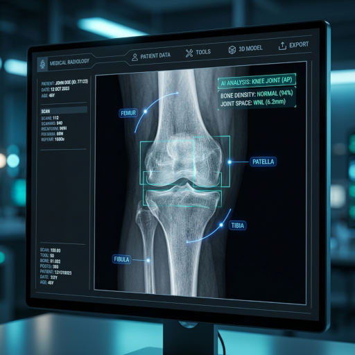
De enkelvork (gevormd door de tibia, fibula en talus) wordt gecontroleerd op symmetrie. Asymmetrie kan wijzen op ligamentair letsel of een fractuur, zelfs als de botten zelf intact lijken.
Calcaneus en Talus (Achtervoet)
Het hielbeen (calcaneus) en de talus worden geëvalueerd op fracturen, hielspoor en standveranderingen die kunnen bijdragen aan hielpijn of instabiliteit.
Middenvoetsbeentjes en Teenkootjes (Voorvoet)
De vijf middenvoetsbeentjes en teenkootjes worden beoordeeld op stressfracturen, dislocatie of hallux valgus-gerelateerde deformiteiten bij het eerste MTP-gewricht.
Zwelling van zacht weefsel en effusie
Omringende zachte weefsels worden geïnspecteerd op zwelling, gewrichtseffusie of vreemde lichamen die gepaard kunnen gaan met trauma of infectie.
Voet- en Enkelröntgenfoto Interpretatie Eenvoudig Gemaakt
De röntgenfoto interpretatietool helpt u de basisbevindingen van voet- en enkelröntgenfoto's te begrijpen met eenvoudige taal, zonder medische kennis te vereisen.
Eenvoudige uitleg voor voet-/enkelröntgenfoto
Ontvang duidelijke, jargonvrije uitleg van wat uw voet- of enkelröntgenfoto onthult.
Educatieve focus op voet-/enkelanatomie
Leer de basisprincipes van voet- en enkelanatomie, fractuurpatronen en veelvoorkomende standafwijkingen.
Gemoedsrust met voet-/enkelröntgenfoto interpretatie
Verminder angst door een basisbegrip te krijgen van uw voet- of enkelröntgenfoto resultaten vóór uw vervolgafspraak.
Hoe de voet- en enkelröntgenfoto interpretatieservice te gebruiken
Vier eenvoudige stappen om een voet- of enkelröntgenfoto interpretatierapport te krijgen via het AI-analysesysteem:
Upload voet- of enkelröntgenfoto
Upload uw voet- of enkelröntgenfoto naar het beveiligde interpretatieplatform. AP-, laterale en oblique opnamen worden allemaal ondersteund.
AI Interpretatie Verwerking
Het AI-systeem analyseert snel de afbeelding en identificeert mogelijke fracturen, gewrichtsveranderingen of standafwijkingen.
Genereer gedetailleerde interpretatie
Het systeem genereert een gemakkelijk te begrijpen interpretatierapport inclusief bevindingen, verklaringen en visuele markeringen.
Bekijk en deel interpretatie
Bekijk de resultaten en deel het rapport optioneel veilig met uw arts of podoloog.
Begrijp uw voet- of enkelröntgenfoto
Upload uw voet- of enkelröntgenfoto om een gemakkelijk te begrijpen uitleg te krijgen
Röntgen, CT, MRI en Echografie
Welke aandoeningen kan een voet- of enkelröntgenfoto detecteren?
Drie-aanzicht (AP, lateraal, mortise) enkelröntgenfoto's en drie-aanzicht (AP, lateraal, oblique) voetröntgenfoto's evalueren de 26 botten van de voet plus de talocrurale en subtalaire gewrichten. Ze zijn het standaard eerstelijns onderzoek na vrijwel elke voet- of enkelblessure.
Enkel (laterale malleolus, mediale malleolus, posterieure malleolus) fracturen
De Weber A/B/C en Lauge-Hansen classificaties die door orthopedisch chirurgen worden gebruikt, zijn direct gebaseerd op de bevindingen van de enkelröntgenfoto — fractuurlocatie, fibula-spiraal en syndesmotische verbreding bepalen chirurgische versus conservatieve zorg.
Fractuur van de basis van het vijfde middenvoetsbeentje (Jones / pseudo-Jones)
Een veelvoorkomende inversieblessure die pijn veroorzaakt aan de laterale middenvoet. De locatie van de fractuurlijn op de röntgenfoto (zone I avulsie versus zone II Jones) bepaalt of gips of chirurgische fixatie nodig is.
Fasciitis plantaris met calcaneusspoor en Achilles-enthesopathie
Een laterale voetröntgenfoto toont het plantaire calcaneusspoor dat wordt gezien bij chronische fasciitis plantaris en de Haglund-deformiteit / posterieure calcaneusspoor geassocieerd met Achilles-tendinopathie.
Lisfranc-letsel (tarsometatarsaal gewrichtscomplex)
Subtiele verbreding tussen het 1e en 2e middenvoetsbeentje (>2 mm) op een gewichtsdragende AP voetröntgenfoto suggereert een Lisfranc-ligamentletsel — een vaak gemiste diagnose met grote langetermijngevolgen indien onbehandeld.
Hallux valgus (bunion) en hallux rigidus
AP gewichtsdragende röntgenfoto's meten de hallux valgus-hoek (HVA) en de intermetatarsale hoek (IMA). Laterale opnamen tonen dorsale osteofyten en gewrichtsspleetvernauwing bij hallux rigidus.
Stressfracturen en Charcot-voet
Middenvoetsbeentje stressfracturen verschijnen vaak pas als subtiele periostale reactie op vervolg-röntgenfoto's. Bij diabetische patiënten documenteren voetröntgenfoto's de botdestructie, fragmentatie en middenvoetcollaps van Charcot-neuroartropathie.
Wanneer moet u een voet- of enkelröntgenfoto laten maken?
De Ottawa Ankle Rules — gevalideerd in meer dan 30 studies — bieden een evidence-based gids voor wanneer een röntgenfoto nodig is na een enkelblessure.
- 1
Enkelpijn plus drukpijn over een van beide malleoli (Ottawa Rules)
Een röntgenfoto is geïndiceerd als de patiënt botgevoeligheid heeft aan de posterieure rand / tip van de laterale of mediale malleolus, of onvermogen om 4 stappen te belasten, zowel direct als op de SEH.
- 2
Middenvoetpijn plus gevoeligheid over het os naviculare of de basis van het 5e middenvoetsbeentje
Deze criteria van de Ottawa Foot Rules begeleiden het aanvragen van voetröntgenfoto's en verminderen onnodige beeldvorming met ~30%.
- 3
Aanhoudende hiel-, voetboog- of voorvoetpijn die langer dan 2 weken aanhoudt
Fasciitis plantaris, Morton's neuroom, stressfractuur of hallux rigidus kunnen allemaal röntgenfoto's vereisen voor evaluatie en chirurgische planning.
- 4
Voetdeformiteit (bunion, klauwtenen, platvoet, Charcot-voet)
Gewichtsdragende röntgenfoto's documenteren de hoeken, gewrichtsbetrokkenheid en het collapspatroon dat nodig is voor het plannen van orthesen of chirurgie.
- 5
Diabetische voetinfectie of niet-genezende ulcus
Röntgenfoto's zijn eerstelijns om onderliggende osteomyelitis (boterosie, periostale reactie) en Charcot-deformiteit bij diabetische patiënten op te sporen.
Voet-/enkelröntgenfoto versus MRI versus CT versus echografie
Elke modaliteit beantwoordt verschillende klinische vragen. Röntgenfoto is bijna altijd het startpunt.
| Beeldvormingsmodaliteit | Het beste in het tonen van | Beperkingen | Kosten & toegang |
|---|---|---|---|
| Voet-/enkelröntgenfoto | Fracturen, stand, artritis, bunions, vreemde lichamen, postoperatieve follow-up | Kan ligamentrupturen, peesletsels, kraakbeen of vroege stressfracturen niet aantonen | Laagste kosten, resultaten in minuten |
| Voet-/enkel-MRI | Achillespeesruptuur, peroneuspeesrupturen, ATFL-ligamentletsel, beenmergoedeem, vroege osteomyelitis | Duur, planningsvertragingen | Hoge kosten |
| Voet-/enkel-CT | Complexe calcaneus-/talusfracturen, chirurgische planning, tarsale coalitie | Hogere straling; beperkte details van zacht weefsel | Midden-hoge kosten |
| Voet-/enkel-echografie | Achilles-tendinopathie, verdikking van de fascia plantaris, Morton's neuroom, ganglioncysten | Operatorafhankelijk; kan geen beenmerg zien | Lage kosten, geen straling |
Hoe u zich voorbereidt op een voet- of enkelröntgenfoto
Voet- en enkelröntgenfoto's vereisen minimale voorbereiding.
Draag een korte broek, losse broek of een ziekenhuisjas
Kleding moet van de voet en enkel worden verwijderd, dus loszittende broeken maken het bezoek sneller.
Verwijder sokken, enkelbandjes en teenringen
Elk metaal in het röntgenveld kan een fractuurlijn verbergen. Teenringen en enkelbandjes moeten worden afgedaan.
Vraag naar gewichtsdragende opnamen als u kunt staan
Gewichtsdragende röntgenfoto's zijn essentieel voor het diagnosticeren van platvoet, hallux valgus, Lisfranc-letsel en het beoordelen van de voetboogstand. Vertel de technicus altijd of u gewicht kunt dragen.
Breng eerdere röntgenfoto's of DICOM-bestanden mee
Vergelijkingsfilms helpen subtiele veranderingen in artritis, botgenezing of de positie van hardware op te sporen.
Beperkingen van AI voet- en enkelröntgenfoto interpretatie
AI markeert veelvoorkomende bevindingen, maar verschillende letsels vereisen aanvullende beeldvorming of specialistisch onderzoek.
- Ligamentletsels zijn onzichtbaar op röntgenfoto's: ATFL-, CFL-, deltoïde- en Lisfranc-ligamentrupturen vereisen MRI. Een normale röntgenfoto en AI-rapport sluiten een hooggradig ligamentletsel niet uit.
- Vroege stressfracturen kunnen worden gemist: Middenvoetsbeentje en naviculare stressfracturen verschijnen vaak pas na 2-3 weken genezing op röntgenfoto's. MRI is de gouden standaard voor vroege detectie.
- Subtiele Lisfranc-letsels vereisen deskundige beoordeling: Een verbreding van 1-2 mm tussen het 1e en 2e middenvoetsbeentje kan duiden op een ernstig ligamentletsel. Bevestig AI-vermoeden altijd met een voet- en enkelspecialist.
CT Read AI voet- en enkelröntgenfoto interpretatie wordt aangeboden voor educatief gebruik. Behandelbeslissingen moeten worden genomen door een gekwalificeerde clinicus.