Interpretazione AI di radiografie di mano e polso
Ottieni aiuto per capire cosa mostra la tua radiografia di mano o polso in un linguaggio semplice. Spiegazioni chiare delle ossa carpali, metacarpi, falangi e articolazioni, inclusi possibili fratture, artrite o problemi di allineamento.
Aiutiamo migliaia di persone a comprendere i risultati delle loro radiografie di mano e polso
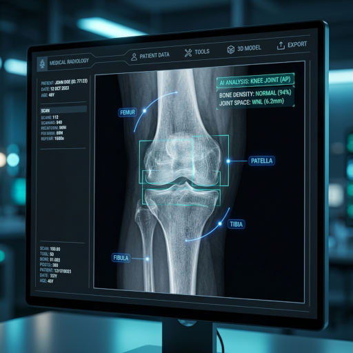
Questo strumento identifica le strutture chiave nelle radiografie di mano e polso e fornisce semplici spiegazioni per aiutarti a capire cosa stai vedendo. Carica la tua radiografia per provarlo tu stesso.
Cos'è un'interpretazione di radiografia di mano e polso?
Una radiografia di mano o polso è un esame di imaging rapido e a bassa radiazione che visualizza le ossa delle dita, del palmo e del polso. CT Read ti aiuta a comprendere le ossa carpali, i metacarpi, le falangi e gli spazi articolari in un linguaggio chiaro e facile da capire.
Ossa carpali (polso)
Vengono valutate le otto piccole ossa carpali del polso, con particolare attenzione allo scafoide, l'osso del polso più comunemente fratturato, spesso non rilevato nelle radiografie iniziali.
Metacarpi e falangi
Le ossa lunghe del palmo (metacarpi) e le ossa delle dita (falangi) vengono valutate per fratture, lussazioni o segni di artrite infiammatoria.
Spazi articolari e segni di artrite
Gli spazi articolari del polso e delle dita vengono controllati per restringimenti, erosioni o speroni ossei che possono indicare osteoartrite, artrite reumatoide o artrite psoriasica.
Gonfiore dei tessuti molli e corpi estranei
I tessuti molli circostanti vengono ispezionati per gonfiore, gas o corpi estranei radio-opachi (ad esempio, vetro o metallo) che possono accompagnare un trauma.
Interpretazione delle radiografie di mano e polso resa semplice
Lo strumento di interpretazione delle radiografie ti aiuta a comprendere i reperti di base delle radiografie di mano e polso utilizzando un linguaggio semplice, senza richiedere alcuna conoscenza medica.
Spiegazioni semplici per radiografie di mano/polso
Ricevi spiegazioni chiare e prive di gergo su ciò che rivela la tua radiografia di mano o polso.
Focus educativo sull'anatomia di mano/polso
Impara le basi dell'anatomia carpale, degli spazi articolari e dei modelli di frattura comuni per discutere meglio i risultati con il tuo medico.
Tranquillità con l'interpretazione della radiografia di mano/polso
Riduci l'ansia acquisendo una comprensione di base dei risultati della tua radiografia di mano o polso in attesa di un parere clinico.
Come utilizzare il servizio di interpretazione delle radiografie di mano e polso
Quattro semplici passaggi per ottenere un referto di interpretazione della radiografia di mano o polso tramite il sistema di analisi AI:
Carica la radiografia di mano o polso
Carica l'immagine della tua radiografia di mano o polso sulla piattaforma di interpretazione sicura. Sono supportate tutte le proiezioni PA, oblique e laterali.
Elaborazione dell'interpretazione AI
Il sistema AI analizza rapidamente l'immagine, identificando potenziali fratture, segni di artrite o anomalie di allineamento.
Genera un'interpretazione dettagliata
Il sistema genera un referto di interpretazione facile da capire che include reperti, spiegazioni e marcatori visivi.
Visualizza e condividi l'interpretazione
Visualizza i risultati e, facoltativamente, condividi il referto in modo sicuro con il tuo medico o terapista.
Comprendi la tua radiografia di mano o polso
Carica l'immagine della tua radiografia di mano o polso per ottenere una spiegazione facile da capire
Raggi X, TC, RMN e Ecografia
Cosa può rilevare una radiografia di mano o polso?
Le proiezioni PA (posteroanteriore), oblique e laterali della mano e del polso consentono una valutazione dettagliata di tutte le 27 ossa, degli spazi articolari e dei tessuti molli circostanti. È l'imaging di prima linea per traumi alla mano, artrite e molte condizioni congenite.
Fratture del radio distale (di Colles)
La frattura più comune negli adulti, solitamente da una caduta su una mano estesa (FOOSH). La radiografia mostra angolazione dorsale, accorciamento del radio e coinvolgimento dello stiloide ulnare che guidano l'ingessatura rispetto alla fissazione chirurgica.
Fratture dello scafoide
Lo scafoide è l'osso carpale più comunemente fratturato e uno dei più spesso non rilevati. Cerca l'interruzione corticale a livello del "collo"; se la radiografia è negativa ma il sospetto clinico persiste, la risonanza magnetica o la ripetizione della radiografia a 10-14 giorni è essenziale a causa del rischio di necrosi avascolare.
Frattura del pugile (collo del 5° metacarpo)
Comune dopo un pugno a pugno chiuso, con angolazione volare della testa del metacarpo. Il grado di angolazione determina se è necessaria la riduzione chiusa o la fissazione chirurgica.
Artrite reumatoide (AR)
La radiografia mostra restringimento simmetrico dello spazio articolare delle articolazioni MCP e PIP, erosioni marginali, osteopenia periarticolare e deviazione ulnare. Il punteggio di Sharp/van der Heijde viene calcolato dalle radiografie PA della mano.
Osteoartrite della base del pollice (OA CMC) e delle articolazioni DIP
L'articolazione CMC del 1° dito e le articolazioni DIP (noduli di Heberden) sono le più comunemente colpite dall'OA della mano. La radiografia mostra restringimento dello spazio articolare, osteofiti e sublussazione della base del pollice.
Corpi estranei e reperti dei tessuti molli
Frammenti di vetro, metallo e ghiaia sono radio-opachi e facilmente visibili. Gonfiore dei tessuti molli, gas e calcificazioni (ad esempio, malattia da pirofosfato di calcio) sono anche documentati sulle radiografie semplici.
Quando è necessaria una radiografia di mano o polso?
Le radiografie di mano/polso vengono richieste per quasi ogni lesione acuta, dolore articolare persistente o sospetta artrite sistemica che colpisce le mani.
- 1
Trauma acuto — cadute, infortuni sportivi o da pugno
Se gonfiore, deformità, lividi o dolorabilità su un osso seguono un trauma, viene richiesta una radiografia per escludere fratture, lussazioni o avulsioni legamentose.
- 2
Dolore persistente al polso, specialmente nella tabacchiera anatomica
La dolorabilità della tabacchiera dopo una FOOSH è preoccupante per una frattura dello scafoide e giustifica l'imaging anche se la radiografia iniziale è negativa.
- 3
Dolore articolare cronico, gonfiore o rigidità mattutina
Il dolore alla mano bilaterale e simmetrico con rigidità che dura più di un'ora al mattino suggerisce artrite infiammatoria (AR o psoriasica). Le radiografie PA della mano fanno parte dell'iter diagnostico.
- 4
Diminuzione della forza di presa o deformità visibile
Deformità a boutonnière, a collo di cigno, deviazione ulnare o sublussazione della base del pollice sono documentate sulla radiografia per classificare la gravità e pianificare l'applicazione di tutori o l'intervento chirurgico.
- 5
Sospetto corpo estraneo dopo un taglio o una puntura
Le radiografie rilevano praticamente tutti i frammenti di metallo e vetro e sono di routine prima dell'esplorazione della ferita.
Radiografia di mano/polso vs risonanza magnetica vs TC vs ecografia
La radiografia è veloce, economica ed eccellente per le ossa, ma le domande sui tessuti molli richiedono un imaging diverso.
| Modalità di imaging | Migliore per mostrare | Limitazioni | Costo e accesso |
|---|---|---|---|
| Radiografia di mano/polso | Fratture, lussazioni, classificazione dell'artrite, allineamento osseo, corpi estranei | Non rileva molte lesioni legamentose, lesioni del TFCC, AR precoce e piccole fratture dello scafoide | Costo più basso, disponibile in qualsiasi pronto soccorso o centro di cure urgenti |
| Risonanza magnetica di mano/polso | Lesioni del legamento scafolunato, lesioni del TFCC, sinovite precoce da AR, edema del midollo osseo, fratture occulte | Costosa; tempo di scansione lungo; non sempre disponibile urgentemente | Costo elevato; tempi di attesa più lunghi |
| TC di mano/polso | Fratture carpali complesse, fratture intra-articolari, pianificazione chirurgica, sospetta pseudoartrosi | Radiazione più elevata; nessun dettaglio dei tessuti molli | Costo medio-alto |
| Ecografia di mano/polso | Lesioni tendinee, cisti gangliari, sinovite da AR, iniezioni guidate | Dipendente dall'operatore; non può valutare l'osso profondo | Basso costo, nessuna radiazione |
Come prepararsi per una radiografia di mano o polso
Non è richiesta alcuna preparazione speciale. Alcuni piccoli accorgimenti miglioreranno la qualità dell'immagine e l'accuratezza dell'interpretazione AI.
Rimuovere tutti i gioielli, orologi e anelli
Gli oggetti metallici appaiono di un bianco brillante sulle radiografie e possono nascondere reperti importanti. Alcuni centri di imaging accettano nastro adesivo temporaneo sugli anelli se la rimozione è impossibile a causa del gonfiore.
Informare il tecnico su precedenti fratture o interventi chirurgici
Placche, viti, fili di Kirschner e vecchie fratture guarite possono mimare una lesione acuta. Conoscere l'anamnesi consente al radiologo (e all'AI) di concentrarsi su nuovi reperti.
Posizionare la mano esattamente come indicato
Le proiezioni standard PA, oblique e laterali richiedono un posizionamento preciso. Una rotazione impropria può nascondere una frattura dello scafoide o distorcere le misurazioni dello spazio articolare.
Portare i file DICOM per l'analisi AI
CT Read produce la migliore interpretazione quando viene fornito il file DICOM originale anziché uno screenshot, perché la risoluzione in scala di grigi e il contrasto della finestra ossea vengono preservati.
Limitazioni dell'interpretazione AI delle radiografie di mano e polso
L'AI è intesa come strumento di triage e formazione. Diverse patologie comuni sono intrinsecamente difficili o impossibili da rilevare con le radiografie semplici.
- Fratture occulte dello scafoide: Fino al 30% delle vere fratture dello scafoide non sono visibili nelle radiografie del primo giorno. Chiunque abbia dolorabilità della tabacchiera dovrebbe essere immobilizzato e riesaminato a 10-14 giorni, indipendentemente dall'output dell'AI.
- Lesioni legamentose e tendinee: La dissociazione scafolunata, le lesioni del TFCC e le rotture tendinee spesso richiedono risonanza magnetica o ecografia; la radiografia e l'AI possono apparire "normali" in loro presenza.
- Artrite infiammatoria precoce: Le erosioni ossee dell'AR possono impiegare 6-12 mesi per apparire sulla radiografia. Un referto AI normale non esclude l'AR precoce — l'esame clinico e gli esami di laboratorio (anti-CCP, RF) rimangono essenziali.
L'interpretazione AI delle radiografie di mano e polso di CT Read è solo a scopo educativo. Confermare sempre qualsiasi reperto anomalo con un chirurgo della mano o un radiologo qualificato.