AI Borst Echografie Interpretatie
Krijg hulp bij het begrijpen van wat uw borst echografie toont in duidelijke taal. Duidelijke uitleg van eenvoudige cysten, complexe massa's en BI-RADS classificatie.
Duizenden vrouwen helpen hun borst echografie resultaten te begrijpen
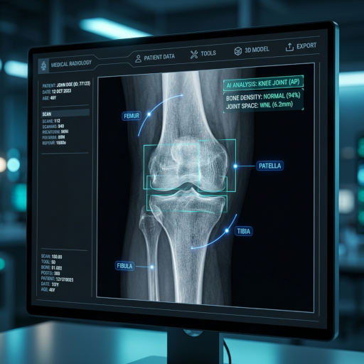
Deze tool analyseert borst echografie beelden en geeft eenvoudige uitleg over cysten, solide massa's en BI-RADS categorisering.
Wat is een borst echografie interpretatie?
Borst echografie is een stralingsvrij beeldvormend onderzoek dat geluidsgolven gebruikt om solide borstmassa's te onderscheiden van met vocht gevulde cysten. CT Read helpt u te begrijpen wat cyste types, kenmerken van solide laesies en BI-RADS categorieën betekenen.
Eenvoudige cysten versus complexe cysten
Eenvoudige cysten zijn anechoïsche, ronde, goed gedefinieerde vochtcollecties (meestal goedaardig, BI-RADS 2). Complexe cysten kunnen debris, septa of solide componenten bevatten en vereisen nader onderzoek.
Kenmerken van solide massa's
Solide massa's worden beoordeeld op vorm, marges, oriëntatie (parallel versus langer-dan-breed) en posterieure akoestische kenmerken. Verdachte kenmerken zijn onregelmatige marges, niet-parallelle oriëntatie en posterieure schaduwvorming.
Vascularisatie (Doppler)
Kleuren-Doppler toont de bloedstroom binnen een laesie. Verhoogde interne vascularisatie kan duiden op een zorgwekkendere laesie, maar wordt geïnterpreteerd samen met de morfologie.
Axillaire lymfeklieren
Axillaire lymfeklieren worden beoordeeld op corticale verdikking, verlies van de vette hilus en asymmetrische grootte – kenmerken die kunnen wijzen op lymfeklierbetrokkenheid.
Borst echografie interpretatie gemakkelijk gemaakt
De echografie interpretatie tool helpt u de basis bevindingen van borst echografie in duidelijke taal te begrijpen.
Eenvoudige uitleg voor borst echografie
Ontvang duidelijke, jargonvrije uitleg over cyste types, solide massa's en BI-RADS categorieën.
Educatieve focus op borstbeeldvorming
Leer de basisprincipes van borst echografie kenmerken en hoe deze zich vertalen naar BI-RADS rapporten.
Gemoedsrust na screening
Verminder angst door borst echografie resultaten te begrijpen vóór uw vervolgafspraak.
Hoe de borst echografie interpretatie service te gebruiken
Vier eenvoudige stappen om een borst echografie interpretatie rapport te krijgen:
Upload borst echografie beelden
Upload één of meer borst echografie beelden (DICOM, JPG of PNG) naar het beveiligde platform.
AI interpretatie verwerking
De AI analyseert de vorm, echogeniciteit en vascularisatie van de laesie en geeft BI-RADS-conforme beschrijvingen.
Genereer gedetailleerde interpretatie
Een gemakkelijk te begrijpen borst echografie interpretatie rapport wordt gegenereerd met uitleg van de belangrijkste bevindingen.
Bekijk en deel interpretatie
Bekijk de resultaten en deel ze optioneel veilig met uw arts of borstspecialist.
Begrijp uw borst echografie
Upload uw borst echografie beeld voor een gemakkelijk te begrijpen uitleg
Röntgen, CT, MRI en Echografie
Welke aandoeningen kan een borst echografie opsporen?
Borst echografie gebruikt hoogfrequente geluidsgolven om borstweefsel af te beelden zonder straling. Het is het eerstelijns beeldvormend onderzoek voor vrouwen onder de 30, voor het evalueren van palpabele knobbels, en als aanvulling op mammografie bij vrouwen met dichte borsten.
Eenvoudige borstcysten
Echografie is de gouden standaard voor het onderscheiden van met vocht gevulde cysten (die bijna altijd goedaardig zijn) van solide massa's. Een eenvoudige cyste – anechoïsch, goed omschreven, met posterieure akoestische versterking – behoeft geen biopsie.
Fibroadenomen
De meest voorkomende goedaardige solide borsttumor bij jonge vrouwen, fibroadenomen verschijnen als ovale, hypoechoïsche massa's met gladde randen en parallelle oriëntatie. De meeste kunnen veilig worden gemonitord zonder biopsie.
Invasieve borstkanker
Verdachte echografie kenmerken zijn onder andere een onregelmatige vorm, niet-parallelle oriëntatie ("langer dan breed"), spiculated of hoekige marges, posterieure schaduwvorming en verhoogde interne vascularisatie op Doppler.
Aanvullende screening voor dichte borsten
Bij vrouwen met extreem dichte borsten (BI-RADS dichtheid d) spoort aanvullende echografie na mammografie 3-4 extra kankers op per 1.000 vrouwen – belangrijk wanneer MRI niet beschikbaar is.
Lactatie- en post-partumproblemen
Echografie diagnosticeert gemakkelijk galactoceles, verstopte melkkanalen, mastitis-gerelateerde abcessen, en is het eerstelijns onderzoek voor zwangere vrouwen of vrouwen die borstvoeding geven met borstklachten.
Lymfeklierbeoordeling
Echografie evalueert axillaire lymfeklieren op verdachte kenmerken (corticale verdikking >3 mm, verlies van vette hilus) en begeleidt fijne-naald aspiratie bij het stadiëren van een bekende kanker.
Wanneer wordt een borst echografie aanbevolen?
Borst echografie is in veel specifieke situaties het juiste eerste beeldvormend onderzoek.
- 1
Palpabele borstknobbel bij elke vrouw
Echografie is eerstelijns voor vrouwen onder de 30 (omdat mammografie beperkt nut heeft bij dichte borsten van jonge vrouwen) en wordt toegevoegd aan mammografie bij oudere vrouwen.
- 2
Zwangere vrouwen of vrouwen die borstvoeding geven met borstsymptomen
Echografie heeft geen straling en is veilig tijdens zwangerschap en borstvoeding, waardoor het de voorkeur heeft als initiële test voor elke borstklacht.
- 3
Aanvullende screening voor vrouwen met dichte borsten
Echografie van de hele borst wordt in veel regio's aanbevolen voor vrouwen met dichte borsten (BI-RADS dichtheid c/d) wanneer MRI niet beschikbaar of betaalbaar is.
- 4
Karakterisering van een mammografische bevinding
Echografie wordt gebruikt om te bepalen of een mammografische massa cysteus of solide is, om de vorm en marges te beoordelen, en om biopsie te begeleiden.
- 5
Beeldgeleide biopsie en procedures
Echografie is de meest voorkomende geleidingsmethode voor borstcorebiopsieën, cyste-aspiraties, markerplaatsingen en pre-operatieve draadlokalisaties.
Borst echografie versus mammografie versus MRI
Elke borstbeeldvormingsmodaliteit heeft unieke voordelen – ze worden vaak samen gebruikt.
| Beeldvormingsmodaliteit | Best in het tonen van | Beperkingen | Kosten & toegang |
|---|---|---|---|
| Borst echografie | Cysten versus solide; palpabele massa's; zwangere/jonge vrouwen; dichte borsten; biopsie begeleiding | Kan calcificaties niet detecteren; lagere gevoeligheid voor DCIS; operatorafhankelijk | Lage kosten, geen straling |
| Mammografie (2D) | Populatiescreening 40+; microcalcificaties; langetermijnvergelijking | Lagere gevoeligheid bij dichte borsten; omvat straling | Lage kosten; breed beschikbaar |
| Tomosynthese (3D mammogram) | Verbeterde gevoeligheid bij dichte borsten; lager recallpercentage | Iets hogere dosis dan 2D; nog niet universeel | Lage-middelmatige kosten |
| Borst MRI | Hoogste gevoeligheid voor invasieve kanker; screening met hoog risico; stadiëring | Duur; vereist IV contrast; lange scantijd; meer vals-positieven | Hoogste kosten |
Hoe u zich voorbereidt op een borst echografie
Borst echografie is een van de gemakkelijkste beeldvormende onderzoeken – geen vasten, geen injecties, geen straling.
Draag een tweedelige outfit
U kleedt zich uit vanaf de taille en trekt een ziekenhuisjas aan. Een tweedelige outfit maakt het bezoek sneller.
Vermijd lotion, deodorant of poeder op de borst of onder de arm
Deze producten kunnen de koppeling van de echografiegel en de beeldkwaliteit beïnvloeden.
Breng eerdere beeldvormings- en biopsierapporten mee
Vergelijking met eerdere mammogrammen, echografieën of MRI is cruciaal voor een accurate interpretatie, vooral als een bekende laesie wordt gevolgd.
Wees voorbereid om een specifiek aandachtsgebied aan te wijzen
Als u kwam voor een palpabele knobbel, markeer het gebied dan vóór het onderzoek. Gerichte scanning van door de patiënt geïdentificeerde bevindingen verbetert de kankerdetectie.
Beperkingen van AI borst echografie interpretatie
Echografie is operatorafhankelijk en heeft inherente beperkingen.
- Kan microcalcificaties niet detecteren: Een belangrijk teken van DCIS – geclusterde microcalcificaties – is onzichtbaar op echografie. Mammografie blijft essentieel voor het screenen van vrouwen boven de 40.
- Operator- en apparatuurafhankelijk: De beeldkwaliteit varieert dramatisch met de ervaring van de echografist en de kwaliteit van de probe. AI-interpretatie is slechts zo goed als het invoerbeeld.
- Vals-positieven leiden tot aanvullende biopsieën: Veel BI-RADS 3 en 4A laesies op echografie blijken goedaardig te zijn bij biopsie. AI helpt het risico te stratificeren, maar elimineert de noodzaak van weefseldiagnose niet.
CT Read AI borst echografie interpretatie is uitsluitend bedoeld voor educatief gebruik. Alle klinische behandeling van borstlaesies moet worden geleid door een gekwalificeerde borstspecialist of radioloog.