KI-gestützte Wirbelsäulen-CT-Befundung
Erhalten Sie Hilfe, um in einfacher Sprache zu verstehen, was Ihr Wirbelsäulen-CT-Scan zeigt. Detaillierte Auswertung der Hals-, Brust- und Lendenwirbel, Bandscheibenräume, Spinalkanal und umgebendes Weichgewebe.
Tausenden von Menschen helfen, ihre Wirbelsäulen-CT-Ergebnisse zu verstehen
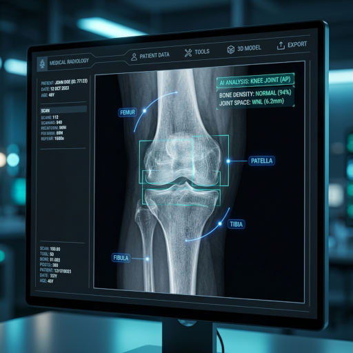
Dieses Tool analysiert Wirbelsäulen-CT-Bilder und liefert klare Erklärungen zu Wirbeln, Bandscheibenräumen, dem Spinalkanal und eventuellen Frakturen oder degenerativen Veränderungen.
Was ist eine Wirbelsäulen-CT-Befundung?
Ein Wirbelsäulen-CT-Scan liefert detaillierte Querschnittsbilder der Hals-, Brust- oder Lendenwirbelsäule. CT Read hilft Ihnen, Wirbelkörper, Bandscheibenräume, die Dimensionen des Spinalkanals zu verstehen und Frakturen oder degenerative Veränderungen zu erkennen.
Wirbelkörperanalyse
Jeder Wirbelkörper wird auf Kompressionsfrakturen, Berstungsfrakturen, lytische oder sklerotische Läsionen und Knochendichte untersucht, die auf Osteoporose hinweisen können.
Bandscheibenräume und degenerative Veränderungen
Bandscheibenhöhen, Vakuumphänomen, Verkalkungen sowie Bandscheibenvorwölbungen oder -vorfälle werden beurteilt. Bandscheiben-Osteophyten-Komplexe, die zu Nervenkompressionen beitragen, können beurteilt werden.
Spinalkanal und Foramina
Der Spinalkanal und die Neuroforamina werden auf Stenosen gemessen. Knöcherne Verengungen können das Rückenmark oder die Nervenwurzeln komprimieren und radikuläre Symptome erklären.
Spondylolisthesis und Ausrichtung
Die Wirbelsäulenausrichtung wird auf Gleiten (Spondylolisthesis), Facettengelenkveränderungen und abnormale Krümmungen wie Skoliose oder Kyphose überprüft.
Wirbelsäulen-CT-Befundung leicht gemacht
Das CT-Befundungstool hilft Ihnen, grundlegende Wirbelsäulen-CT-Befunde in einfacher Sprache zu verstehen.
Einfache Erklärungen für Wirbelsäulen-CT
Erhalten Sie klare, jargonfreie Erklärungen zu Wirbeln, Bandscheiben und dem Spinalkanal.
Bildungsschwerpunkt Wirbelsäulenanatomie
Lernen Sie die Grundlagen der Wirbelsäulenanatomie und häufige CT-Befunde wie Kompressionsfrakturen und Stenosen.
Beruhigung vor dem Facharztbesuch
Reduzieren Sie Ängste, indem Sie die Wirbelsäulen-CT-Ergebnisse verstehen, bevor Sie sie mit einem Wirbelsäulenchirurgen oder Neurologen besprechen.
So nutzen Sie den Wirbelsäulen-CT-Befundungsdienst
Vier einfache Schritte, um einen Wirbelsäulen-CT-Befundbericht zu erhalten:
Wirbelsäulen-CT-Bilder hochladen
Laden Sie eine oder mehrere Wirbelsäulen-CT-Schichten (DICOM, JPG oder PNG) auf die sichere Plattform hoch.
KI-Interpretationsverarbeitung
Die KI analysiert Wirbel, Bandscheiben und den Spinalkanal auf Frakturen, Stenosen und degenerative Veränderungen.
Detaillierte Interpretation erstellen
Ein leicht verständlicher Wirbelsäulen-CT-Befundbericht wird mit Erklärungen der wichtigsten Befunde erstellt.
Interpretation ansehen und teilen
Sehen Sie sich die Wirbelsäulen-CT-Ergebnisse an und teilen Sie sie optional sicher mit Ihrem Arzt.
Verstehen Sie Ihren Wirbelsäulen-CT-Scan
Laden Sie Ihr Wirbelsäulen-CT-Bild hoch, um eine leicht verständliche Erklärung zu erhalten
Röntgen, CT, MRT und Ultraschall
Welche Erkrankungen kann ein Wirbelsäulen-CT erkennen?
Das Wirbelsäulen-CT liefert Querschnittsbilder der Hals-, Brust- und Lendenwirbel sowie der umgebenden paraspinalen Strukturen. Mit multiplanaren Reformationen und 3D-Rekonstruktionen ist das CT der Goldstandard für die Beurteilung der knöchernen Wirbelsäule und komplexer Traumata.
Wirbelfrakturen und Trauma
CT ist die bildgebende Untersuchung der ersten Wahl bei Wirbelsäulentraumata und ist wesentlich empfindlicher als Röntgenaufnahmen für Kompressions-, Berstungs-, Chance-, Querfortsatz- und Densfrakturen. Die von Wirbelsäulenchirurgen verwendete AOSpine-Klassifikation basiert direkt auf CT-Befunden.
Spinalkanalstenose und degenerative Veränderungen
CT zeigt Facettengelenkhypertrophie, Ligamentum-flavum-Verkalkung, Bandscheibenraumverengung und knöcherne Foramenstenose. CT-Myelographie ergänzt MRT bei Patienten mit Implantaten.
Bandscheibenvorfall (insbesondere verkalkt)
Während MRT bei weichen Bandscheibenvorfällen bevorzugt wird, zeigt CT verkalkte Bandscheibenvorfälle hervorragend und kann akute Bandscheibenvorwölbungen bei Patienten zeigen, die kein MRT erhalten können.
Spondylolyse und Spondylolisthesis
CT zeigt deutlich Defekte der Pars interarticularis (Spondylolyse) – die häufigste Ursache für Kreuzschmerzen bei jugendlichen Sportlern – und klassifiziert das Wirbelgleiten (Meyerding I–V).
Wirbelsäulentumoren und Metastasen
CT erkennt lytische und blastiche Knochenmetastasen, primäre Knochentumoren (Osteoidosteom, Chordom) und pathologische Frakturen. Es wird auch zur Planung von Biopsien und Strahlentherapie eingesetzt.
Postoperative Hardware- und Fusionsbeurteilung
CT ist die bildgebende Untersuchung der Wahl nach Wirbelsäulenversteifungsoperationen, um die Position der Pedikelschrauben, die Bildung der knöchernen Fusionsmasse zu beurteilen und eine Lockerung oder Bruch der Hardware auszuschließen.
Wann wird ein Wirbelsäulen-CT angeordnet?
Wirbelsäulen-CT wird Röntgenaufnahmen bei fast allen mittelschweren bis schweren Wirbelsäulenpathologien und MRT vorgezogen, wenn knöcherne Details Priorität haben.
- 1
Bedeutendes Wirbelsäulentrauma oder Polytrauma
CT ist die Erstuntersuchung bei Hals-, Brust- und Lendenwirbelsäulenverletzungen basierend auf den kanadischen C-Spine- und NEXUS-Kriterien.
- 2
Plötzliche starke Rückenschmerzen mit neurologischen Symptomen
Sattelanästhesie, Harnverhalt oder fortschreitende Schwäche erfordern eine dringende Bildgebung – CT kann schnell Frakturen, Tumoren oder Blutungen ausschließen, wenn MRT nicht verfügbar ist.
- 3
Präoperative Planung für Wirbelsäulenchirurgie
CT liefert die knöchernen Details, die für die Planung von Pedikelschrauben, die Korrektur von Deformitäten und die Tumorresektion erforderlich sind.
- 4
Verdacht auf Spondylolyse bei jungen Sportlern
Ein jugendlicher Turner oder Fußballspieler mit anhaltenden Kreuzschmerzen benötigt oft ein CT, um einen Pars-Defekt zu erkennen, den Röntgenaufnahmen möglicherweise übersehen.
- 5
Postoperative Beurteilung
Nach Fusion oder Instrumentierung dokumentiert CT den Fusionsfortschritt, die Hardware-Integrität und eventuelle Komplikationen.
Wirbelsäulen-CT vs. MRT vs. Röntgen vs. Knochenszintigraphie
Jede Modalität hat Stärken. CT ist am besten für Knochen und akute Traumata; MRT ist am besten für Rückenmark und weiche Bandscheiben; Röntgen ist am besten für Screening und Ausrichtung.
| Bildgebende Modalität | Am besten geeignet für die Darstellung von | Einschränkungen | Kosten & Zugang |
|---|---|---|---|
| Wirbelsäulen-CT | Frakturen, knöcherne Stenosen, verkalkte Bandscheiben, Hardware, komplexe Traumata, Fusionsbeurteilung | Höhere Strahlung; begrenzte Weichteildetails; kann das Rückenmark nicht direkt visualisieren | Mittlere Kosten; weit verbreitet in der Notaufnahme |
| Wirbelsäulen-MRT | Rückenmark, Nervenwurzeln, weicher Bandscheibenvorfall, Knochenmarkinfiltration, Infektion, Tumorausdehnung | Lange Scanzeit; teuer; kontraindiziert bei einigen Implantaten und Herzschrittmachern | Hohe Kosten; längere Wartezeit |
| Wirbelsäulen-Röntgen | Ausrichtung, Skoliose-Überwachung, Spondylolisthesis-Graduierung, Trauma-Screening | Übersieht viele Frakturen; kann Rückenmark oder Bandscheibe nicht zeigen | Geringste Kosten; am schnellsten |
| Knochenszintigraphie / SPECT | Aktive Stressfrakturen, Metastasen-Screening, Infektions-Screening | Geringe räumliche Auflösung; unspezifische Befunde | Mittlere Kosten; Spezialabteilung |
So bereiten Sie sich auf ein Wirbelsäulen-CT vor
Ein Wirbelsäulen-CT dauert typischerweise 5–10 Minuten und erfordert minimale Vorbereitung.
Tragen Sie lockere, metallfreie Kleidung
Sie werden möglicherweise gebeten, sich umzuziehen, um Reißverschlüsse, Druckknöpfe und Bügel-BHs zu entfernen. Metallartefakte können die Bildqualität an der Wirbelsäule beeinträchtigen.
Informieren Sie das Personal über eine Schwangerschaft
Ein Wirbelsäulen-CT beinhaltet eine moderate Strahlung (Lendenwirbelsäule ~6 mSv); eine Schwangerschaft ist eine relative Kontraindikation, es sei denn, sie ist absolut notwendig.
Informieren Sie das Personal über Nierenprobleme, falls Kontrastmittel benötigt wird
Die meisten Wirbelsäulen-CTs werden ohne intravenöses Kontrastmittel durchgeführt. Falls Kontrastmittel erforderlich ist (Verdacht auf Tumor, Infektion, Gefäßverletzung), werden aktuelle Nierenfunktionstests überprüft.
Bringen Sie frühere Bilder auf CD oder per DICOM-Übertragung mit
Frühere CT- oder MRT-Bilder verbessern die Genauigkeit der KI und des Radiologen erheblich, indem sie einen direkten Vergleich von Bandscheibenhöhen, Ausrichtung und neuen Läsionen ermöglichen.
Einschränkungen der KI-Wirbelsäulen-CT-Befundung
CT und KI haben auch in erfahrenen Händen wichtige Einschränkungen.
- Kann das Rückenmark nicht direkt visualisieren: Rückenmarksverletzungen, Demyelinisierung, Syringomyelie und intrinsische Rückenmarksläsionen erfordern ein MRT. Ein normales Wirbelsäulen-CT schließt eine Rückenmarkspathologie nicht aus.
- Weicher Bandscheibenvorfall kann subtil sein: Nicht verkalkte Bandscheibenvorfälle sind im MRT besser sichtbar. CT kann kleine weiche Bandscheibenvorfälle, die eine Radikulopathie verursachen, übersehen.
- Höhere Strahlendosis als Röntgen: Ein Lendenwirbelsäulen-CT liefert ~6 mSv (etwa 2 Jahre Hintergrundstrahlung). Das Risiko-Nutzen-Verhältnis muss bei jungen Patienten und während der Schwangerschaft berücksichtigt werden.
Die CT Read KI-Wirbelsäulen-CT-Befundung dient nur zu Bildungs- und Triagezwecken. Alle Behandlungsentscheidungen müssen einen Wirbelsäulenchirurgen, Neurochirurgen oder Radiologen einbeziehen.