KI-Hüft-Röntgenbild-Interpretation
Erhalten Sie Hilfe, um zu verstehen, was Ihr Hüft-Röntgenbild in Alltagssprache zeigt. Klare Erklärungen des Hüftgelenks, des Femurkopfs, des Acetabulums und der umgebenden Knochenstrukturen.
Tausenden von Menschen helfen, ihre Hüft-Röntgenbilder zu verstehen
Dieses Tool identifiziert Schlüsselstrukturen in Hüft-Röntgenbildern und bietet einfache Erklärungen, um Ihnen zu helfen, zu verstehen, was Sie sehen. Laden Sie Ihr Röntgenbild hoch, um es selbst auszuprobieren.
Was ist eine Hüft-Röntgenbild-Interpretation?
Eine Hüft-Röntgenaufnahme ist eine schnelle, schmerzlose bildgebende Untersuchung, die Bilder des Hüftgelenks und der umgebenden Knochen erstellt. CT Read hilft Ihnen, diese Bilder zu verstehen, indem es Ihren Femurkopf, Ihr Acetabulum, Ihren Gelenkspalt und Ihre Knochendichte in einfacher Sprache klar erklärt.
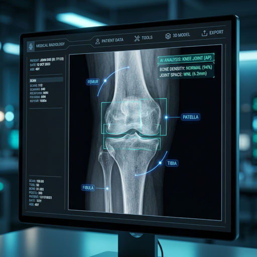
Hüftgelenkspalt
Der Raum zwischen dem Femurkopf (Kugel) und dem Acetabulum (Pfanne) wird auf Verengungen untersucht, die auf Hüftarthrose oder Knorpelverlust hinweisen können.
Femurkopf und -hals
Der kugelförmige obere Teil des Oberschenkelknochens und sein Hals werden auf Frakturen, avaskuläre Nekrose oder Anzeichen eines Impingements untersucht, die Hüftschmerzen verursachen können.
Acetabulum (Hüftpfanne)
Die schalenförmige Pfanne des Beckens wird auf Form, Tiefe und Anzeichen von Dysplasie, Frakturen oder degenerativen Veränderungen untersucht, die die Gelenkstabilität beeinträchtigen.
Knochendichte und Ausrichtung
Knochendichte und die allgemeine Hüftausrichtung werden bewertet, um Osteoporose, Beinlängendifferenz oder Ausrichtungsprobleme zu identifizieren, die zu Symptomen beitragen.
Hüft-Röntgenbild-Interpretation leicht gemacht
Das Röntgenbild-Interpretationstool hilft Ihnen, grundlegende Hüft-Röntgenbefunde in einfacher Sprache zu verstehen, ohne medizinisches Wissen vorauszusetzen.
Einfache Erklärungen zur Hüft-Röntgenbild-Interpretation
Erhalten Sie klare, jargonfreie Erklärungen darüber, was Ihr Hüft-Röntgenbild zeigt, und machen Sie medizinische Informationen für jedermann zugänglich.
Bildungsschwerpunkt auf Hüft-Röntgenbild-Interpretation
Dieses Tool wurde entwickelt, um aufzuklären und zu informieren und Ihnen zu helfen, Ihre Informationen zur Hüftgesundheit besser zu verstehen, bevor Sie sie mit Ihrem Arzt besprechen.
Seelenfrieden mit der Hüft-Röntgenbild-Interpretation
Reduzieren Sie Ängste, indem Sie ein grundlegendes Verständnis Ihrer Hüft-Röntgenbilder erlangen, während Sie darauf warten, mit Ihrem Arzt zu sprechen.
So nutzen Sie den Hüft-Röntgenbild-Interpretationsdienst
Vier einfache Schritte, um einen Hüft-Röntgenbild-Interpretationsbericht über das KI-Analysesystem zu erhalten:
Hüft-Röntgenbild zur Interpretation hochladen
Laden Sie Ihr Hüft-Röntgenbild auf die sichere Interpretationsplattform hoch. Verschiedene gängige Bildformate werden für eine genaue Analyse unterstützt.
KI-Interpretationsverarbeitung
Das KI-System führt schnell eine Hüft-Röntgenbild-Interpretation durch und identifiziert potenzielle Gelenk- und Knochenanomalien.
Detaillierte Interpretation erstellen
Das System erstellt einen leicht verständlichen Hüft-Röntgenbild-Interpretationsbericht mit Befunden, Erklärungen und visuellen Markierungen.
Interpretation anzeigen und teilen
Zeigen Sie die Ergebnisse der Hüft-Röntgenbild-Interpretation an und teilen Sie den Bericht optional sicher mit Ihrem Arzt.
Verstehen Sie Ihr Hüft-Röntgenbild
Laden Sie Ihr Hüft-Röntgenbild hoch, um eine leicht verständliche Erklärung zu erhalten
Röntgen, CT, MRT und Ultraschall
Welche Erkrankungen kann ein Hüft-Röntgenbild erkennen?
Ein standardmäßiges AP- (anteroposterior) und laterales Hüft-Röntgenbild bietet Radiologen eine schnelle, kostengünstige Ansicht der knöchernen Anatomie des Hüftgelenks, des Femurkopfs und -halses, des Acetabulums und des oberen Oberschenkelknochens. Es ist die bildgebende Untersuchung der ersten Wahl für fast jede Hüftbeschwerde bei Erwachsenen und kann die folgenden Erkrankungen identifizieren oder stark nahelegen.
Hüftarthrose (OA)
Gelenkspaltverengung (insbesondere superolateral), subchondrale Sklerose, marginale Osteophyten und subchondrale Zysten sind die vier klassischen Anzeichen einer Hüft-OA auf dem Röntgenbild. Das von Orthopäden verwendete Tönnis-Grading-System (0–3) basiert direkt auf diesen einfachen Röntgenbefunden.
Schenkelhals- und intertrochantäre Frakturen
Hüft-Röntgenbilder bleiben die Standarduntersuchung der ersten Wahl nach einem Sturz bei älteren Erwachsenen. Achten Sie auf kortikalen Stufenbildung, trabekuläre Angulation und Verkürzung / Außenrotation des Beins in der AP-Ansicht. Subtile, nicht dislozierte Frakturen können bei anhaltenden Schmerzen eine MRT-Nachuntersuchung erfordern.
Avaskuläre Nekrose (AVN) des Femurkopfs
Frühe AVN kann subtile subchondrale Transluzenz (das „Sichelzeichen“) zeigen, gefolgt von einer Abflachung und einem Kollaps des Femurkopfs in fortgeschrittenen Stadien. Die Ficat-Klassifikation (I–IV) wird auf einfachen Röntgenbildern eingestuft und leitet Entscheidungen zur Hüftgelenkerhaltung vs. -ersatz.
Entwicklungsbedingte Hüftdysplasie & FAI
Ein flaches Acetabulum, ein lateraler Center-Edge-Winkel (LCEA) unter 25°, ein „unbedeckter“ Femurkopf oder eine nicht-sphärische Kopf-Hals-Verbindung (Cam-Morphologie) deuten auf eine Entwicklungsdysplasie oder ein femoroacetabuläres Impingement hin – häufige Ursachen für Hüftschmerzen bei jungen Erwachsenen.
Beurteilung von Hüftimplantaten / Prothesen
Bei Patienten, die eine totale Hüftendoprothese (THA) erhalten haben, werden Röntgenbilder verwendet, um die Ausrichtung der Komponenten, die Symmetrie der Beinlänge, die periprothetische Transluzenz (Lockerung), heterotope Knochenbildung und periprothetische Frakturen zu beurteilen.
Becken- & Schambeinastfrakturen
AP-Beckenansichten, die zusammen mit dem Hüftfilm aufgenommen werden, können Schambeinastfrakturen, Sakralinsuffizienzfrakturen und eine SI-Gelenkverbreiterung aufzeigen – besonders wichtig bei älteren Patienten nach Stürzen mit geringer Energie.
Wann sollte man ein Hüft-Röntgenbild machen lassen?
Ein Hüft-Röntgenbild ist angebracht, wenn ein Arzt eine knöcherne Ursache für Hüft-, Leisten- oder Oberschenkelschmerzen ausschließen muss. Häufige Szenarien sind:
- 1
Schmerzen oder Hinken nach einem Sturz, insbesondere bei Erwachsenen über 60
Ein Hüft-Röntgenbild ist die erste bildgebende Untersuchung nach fast jedem Sturz bei Erwachsenen mit Schmerzen im Hüftbereich, um eine Schenkelhals- oder intertrochantäre Fraktur auszuschließen, beides chirurgische Notfälle.
- 2
Chronische Leisten- oder Gesäßschmerzen, die sich bei Belastung verschlimmern
Schmerzen beim Stehen, Treppensteigen oder Drehen des Beins sind eine klassische Präsentation von Hüftarthrose oder Labrumpathologie – das Röntgenbild ist der erste Schritt zur Beurteilung des Gelenkspalts und der knöchernen Ausrichtung.
- 3
Eingeschränkter Bewegungsumfang oder Steifheit, insbesondere morgens
Eine verminderte Innenrotation ist eines der frühesten körperlichen Untersuchungszeichen einer Hüft-OA. Einfache Röntgenbilder dokumentieren die zugrunde liegenden knöchernen Veränderungen, die die Steifheit erklären.
- 4
Postoperative Nachsorge nach THA oder Frakturfixation
Routinemäßige Röntgenbilder nach 6 Wochen, 3 Monaten, 1 Jahr und danach jährlich werden empfohlen, um die Implantatposition, die Heilung und periprothetische Knochenveränderungen zu überwachen.
- 5
Pädiatrische oder jugendliche Hüftschmerzen (z. B. Verdacht auf SCFE oder Perthes)
Bei Kindern und Jugendlichen ist ein Froschbein-Seiten-Hüft-Röntgenbild unerlässlich, um eine Epiphyseolysis capitis femoris (SCFE) oder Morbus Legg-Calvé-Perthes auszuschließen, beides zeitkritische Diagnosen.
Hüft-Röntgen vs. MRT vs. CT vs. Ultraschall: Welches benötigen Sie?
Jede bildgebende Modalität beantwortet unterschiedliche klinische Fragen. Verwenden Sie diese Tabelle, um zu verstehen, wann ein Röntgenbild ausreicht und wann eine fortgeschrittenere Bildgebung erforderlich ist.
| Bildgebende Modalität | Am besten geeignet zur Darstellung von | Einschränkungen | Kosten & Zugang |
|---|---|---|---|
| Hüft-Röntgen | Knochenanatomie, Frakturen, Gelenkspalt, Arthrose-Grading, Implantat-Nachsorge | Kann Knorpel, Labrum, Sehnen oder Muskeln nicht zeigen; übersieht 5–10 % okkulter Frakturen | Geringste Kosten, weit verbreitet, Ergebnisse in Minuten |
| Hüft-MRT | Labrumrisse, Knorpelschäden, AVN (frühestes Anzeichen), Knochenmarködem, okkulte Frakturen | Lange Scanzeit (30–60 Min.), teuer, kontraindiziert bei einigen Implantaten | Höchste Kosten, längere Wartezeiten für Termine |
| Hüft-CT | Komplexe Frakturen, präoperative OP-Planung, Beckenringverletzungen, 3D-Knochenrekonstruktion | Höhere Strahlendosis als Röntgen; weniger Details bei Weichteilen | Mittlere bis hohe Kosten, in den meisten Notaufnahmen verfügbar |
| Hüft-Ultraschall | Pädiatrisches DDH-Screening (unter 6 Monaten), Gelenkergüsse, gezielte Injektionen | Operatorabhängig; kann tiefe Knochenanatomie bei Erwachsenen nicht beurteilen | Geringe Kosten, keine Strahlung |
So bereiten Sie sich auf ein Hüft-Röntgenbild vor
Hüft-Röntgenbilder erfordern fast keine Vorbereitung, aber ein paar einfache Schritte können ein klareres Bild und eine genauere KI-Interpretation liefern.
Tragen Sie lockere Kleidung ohne Metall
Metallreißverschlüsse, Knöpfe, Gürtelschnallen und Bügel-BHs können als dichte weiße Flecken erscheinen und knöcherne Details verdecken. Möglicherweise werden Sie gebeten, sich in ein Krankenhauskittel umzuziehen.
Entfernen Sie Schmuck, Münzen und Telefone aus Ihren Taschen
Alles Metallische im Röntgenfeld kann mit einer Anomalie verwechselt werden. Nehmen Sie vor dem Scan Schmuck im Hüftbereich ab und leeren Sie Ihre Taschen.
Informieren Sie den Techniker, wenn Sie schwanger sind oder sein könnten
Obwohl die Strahlendosis eines Hüft-Röntgenbildes gering ist (etwa 0,7 mSv), werden schwangeren Patientinnen in der Regel alternative Bildgebungsverfahren oder Abschirmungen angeboten.
Bringen Sie, falls vorhanden, frühere Bilder mit
Der Vergleich des heutigen Röntgenbildes mit einem früheren Film hilft, subtile Veränderungen (z. B. fortschreitende Arthritis oder Implantatlockerung) zu erkennen. Der KI-Interpreter funktioniert am besten, wenn er mit DICOM-Dateien in voller Auflösung gefüttert wird.
Wichtige Einschränkungen der KI-Hüft-Röntgenbild-Interpretation
Die KI von CT Read ist als Bildungs- und Triage-Tool konzipiert. Sie ist kein Ersatz für einen zertifizierten Radiologen oder Orthopäden. Beachten Sie die folgenden Einschränkungen, bevor Sie sich auf eine Ausgabe verlassen:
- Keine Weichteilbeurteilung: Röntgenbilder – und damit die KI – können Labrumrisse, Knorpelschäden, Tendinopathien oder Muskelverletzungen nicht zuverlässig erkennen. Wenn Ihre Symptome auf diese Erkrankungen hindeuten, ist eine MRT erforderlich.
- Okkulte Frakturen können übersehen werden: Bis zu 10 % der Schenkelhalsfrakturen sind auf den ersten Röntgenbildern nicht sichtbar. Wenn Sie nach einem Sturz anhaltende Schmerzen beim Gehen haben, fragen Sie Ihren Arzt nach einer MRT, auch wenn das Röntgenbild normal aussieht.
- Keine klinische Entscheidungsfindung: Die KI kennzeichnet potenzielle Befunde; sie stuft keine Krankheiten ein, verschreibt keine Behandlungen und bestimmt keine Operationsfähigkeit. Besprechen Sie den Bericht immer mit Ihrem behandelnden Arzt.
Die KI-Hüft-Röntgenbild-Interpretation von CT Read dient der Patientenaufklärung und ist keine medizinische Diagnose. Suchen Sie bei medizinischen Beschwerden immer den Rat eines qualifizierten Gesundheitsdienstleisters.